

Apparent diffusion coefficient and arterial spin labeling perfusion of conventional chondrosarcoma in the parafalcine region: a case report
- PMID: 29487660
- PMCID: PMC5826694
- DOI: 10.1016/j.radcr.2017.09.021
Apparent diffusion coefficient and arterial spin labeling perfusion of conventional chondrosarcoma in the parafalcine region: a case report
Abstract
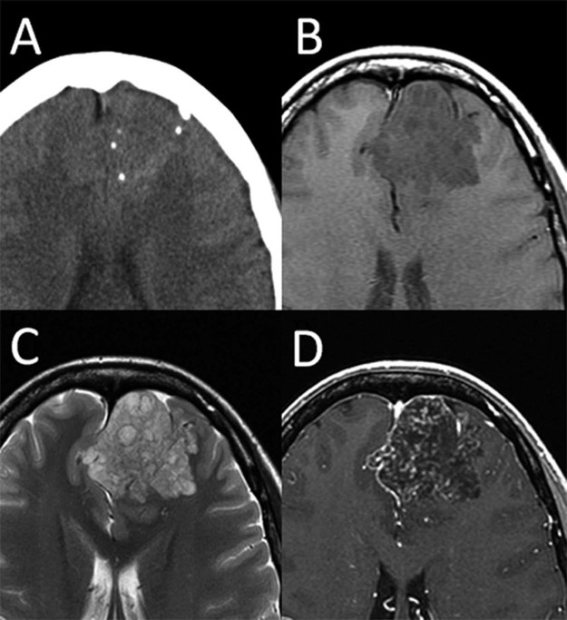
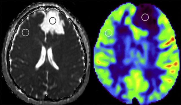
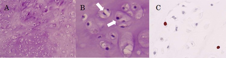
Intracranial chondrosarcoma is a very rare malignant tumor of the central nervous system, and is difficult to preoperatively distinguish from other tumors using conventional imaging techniques. Here, we report the case of a 24-year-old woman who presented with mild headache due to chondrosarcoma in the frontal lobe. Preoperative conventional images showed findings typical of an oligodendroglial tumor. However, high apparent diffusion coefficient (ADC) value and extreme hypoperfusion on arterial spin labeling (ASL) were inconsistent with oligodendroglial tumor characteristics. The tumor was completely removed using a standard surgical procedure. Histologic diagnosis was a conventional (classic) chondrosarcoma. High ADC and hypoperfusion on ASL represented low cellularity and low vascularity within conventional chondrosarcoma, respectively. We discuss the utility of ADC and ASL for the preoperative diagnosis of conventional chondrosarcoma.
Keywords: ADC; ASL; Intracranial chondrosarcoma; MRI.
Figures
Similar articles
-
Intratumor Heterogeneity of Perfusion and Diffusion in Clear-Cell Renal Cell Carcinoma: Correlation With Tumor Cellularity.Clin Genitourin Cancer. 2016 Dec;14(6):e585-e594. doi: 10.1016/j.clgc.2016.04.007. Epub 2016 Apr 22. Clin Genitourin Cancer. 2016. PMID: 27209349 Free PMC article.
-
Intravoxel incoherent motion diffusion-weighted imaging analysis of diffusion and microperfusion in grading gliomas and comparison with arterial spin labeling for evaluation of tumor perfusion.J Magn Reson Imaging. 2016 Sep;44(3):620-32. doi: 10.1002/jmri.25191. Epub 2016 Feb 16. J Magn Reson Imaging. 2016. PMID: 26880230
-
Correlation of biexponential diffusion parameters with arterial spin-labeling perfusion MRI: results in transplanted kidneys.Invest Radiol. 2013 Mar;48(3):140-4. doi: 10.1097/RLI.0b013e318277bfe3. Invest Radiol. 2013. PMID: 23249648 Clinical Trial.
-
Intracranial Nonskull-Based Chondrosarcoma Arising from the Sagittal Sinus: A Case Report and Review of the Literature.World Neurosurg. 2018 Dec;120:234-239. doi: 10.1016/j.wneu.2018.08.239. Epub 2018 Sep 8. World Neurosurg. 2018. PMID: 30205213 Review.
-
Intracranial mesenchymal chondrosarcoma: case report and literature review.Br J Neurosurg. 2012 Dec;26(6):912-4. doi: 10.3109/02688697.2012.697219. Epub 2012 Jun 25. Br J Neurosurg. 2012. PMID: 22731866 Review.
Cited by
-
CT and MRI findings of intracranial extraskeletal mesenchymal chondrosarcoma-a case report and literature review.Transl Cancer Res. 2022 Sep;11(9):3409-3415. doi: 10.21037/tcr-21-2547. Transl Cancer Res. 2022. PMID: 36237268 Free PMC article.
References
-
- Dorfman H.D., Czerniak B. 1998. Bone tumors. St Louis, Baltimore, Boston, Carlsbad, Chicago, Minneapolis, New York, Philadelphia, Portland, London, Milan, Sydney, Tokyo, Toronto: Mosby. Reference to a chapter in an edited book: Benign cartilage lesions.
-
- Antonescu C.R., Paulus W., Perry A., Rushing E.J., Hainfellner J.A., Bouvier C. 2016. Other mesenchymal tumours. In: Louis DN, Ohgaki H, Wiestler OD, et al. editors. WHO Classification of Tumours of the Central Nervous System, Lyon: International Agency for Research on Cancer (IARC) p. 258–264.
-
- Chaskis C., Michotte A., Goossens A., Stadnik T., Koerts G., D'Haens J. Primary intracerebral myxoid chondrosarcoma. Case illustration. J Neurosurg. 2002;97:228. - PubMed
Publication types
LinkOut - more resources
Full Text Sources
Other Literature Sources
