. 2017 Dec 31;9(8):8197-8203.
doi: 10.18632/oncotarget.23793. eCollection 2018 Jan 30.

Complete response in a patient with liver metastases from breast cancer employing hepatic arterial infusion 5-fluorouracil based chemotherapy plus systemic nab-paclitaxel

Affiliations

Complete response in a patient with liver metastases from breast cancer employing hepatic arterial infusion 5-fluorouracil based chemotherapy plus systemic nab-paclitaxel

Girolamo Ranieri et al. Oncotarget. .

Abstract

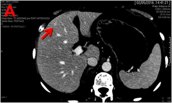
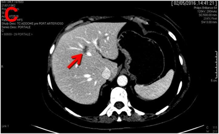
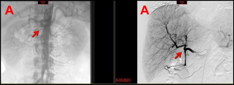
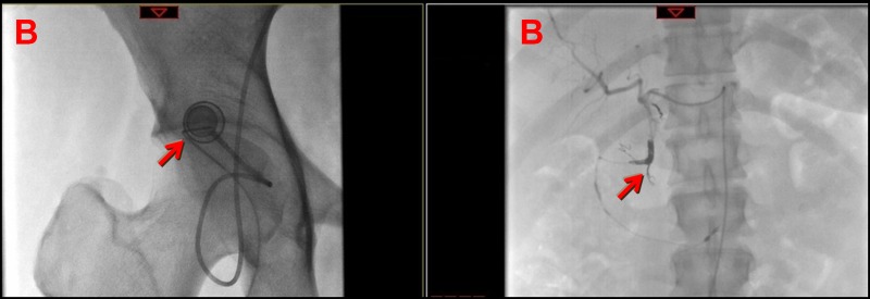
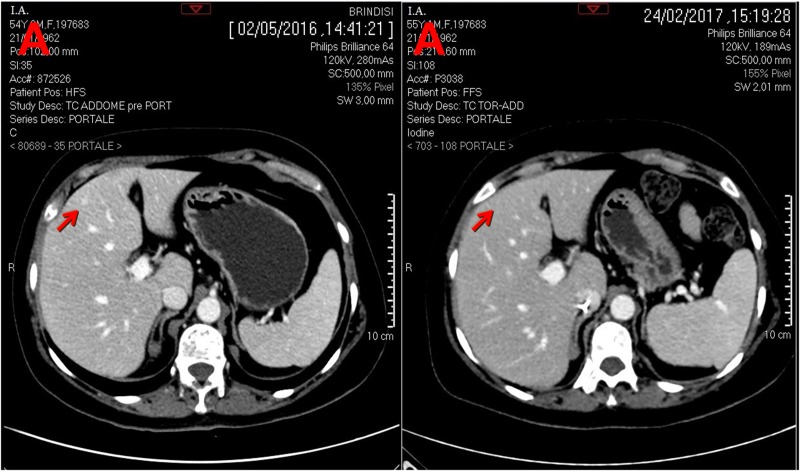
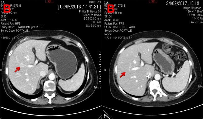
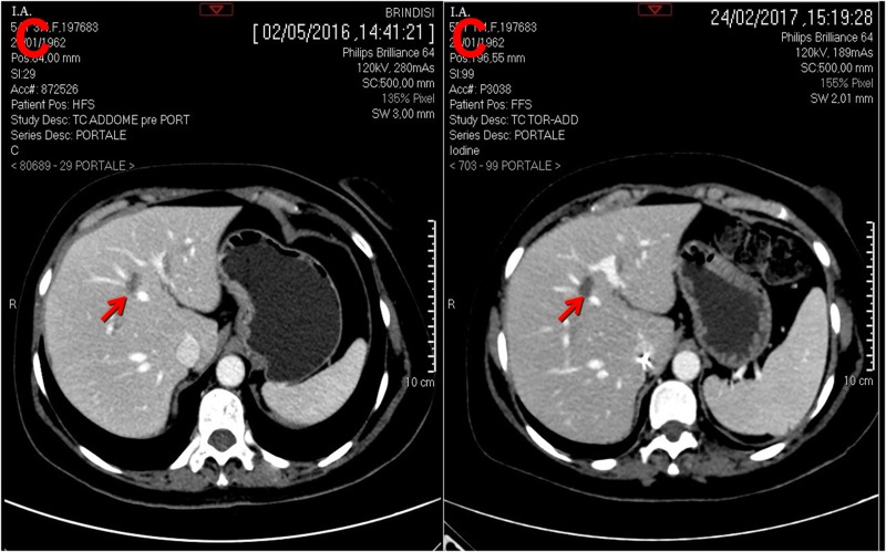
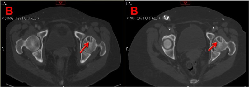
About half of patients with metastatic breast cancer (mBC) have unresectable liver metastases (LMs) or liver-predominant disease (LPD). Unfortunately systemic chemotherapy has limited tumor response due to LMs are supplied by hepatic artery. Hepatic intra-arterial (HAI) have antitumor activity in pretreated patients with LMs. Here we report the case of a 55-year-old woman affected by BCLPD and heavily pretreated. LMs responded to treatment based on HAI with 5-fluorouracil and nab-paclitaxel systemic chemotherapy, and they completely disappeared on a CT-scan. We conclude that this combination chemotherapy is safe and may be very useful for the treatment of patients with BCLPD. Therefore, this combination should be evaluated in a large study.

Keywords: breast cancer; hepatic intra-arterial chemotherapy; liver metastases; nab-paclitaxel.


Conflict of interest statement

CONFLICTS OF INTEREST The authors confirm that there are no conflicts of interest.

Figures

Figure 1
Figure 1. Computed tomography (CT) scan performed on April 2016 shows three liver lesions (LLs) in arterial phase
a. The arrow indicates LL (diameter: 7 mm) with contrast enhancement suspected of metastasis at 4th segment; b. The arrow indicates LL (diameter: 19 mm) with contrast enhancement suspected of metastasis at 5th segment; c. The arrow indicates LL (diameter: 18 mm) without contrast enhancement, doubtful lesion for metastasis at 8th-4th segment and in differential diagnosis with angioma.
Figure 2
Figure 2. Arterial phase of contrast angiogram performed before (a) and after (b) the placement of the subcutaneous port and of the hepatic-artery catheter
a. Arterial phase of contrast angiogram shows the celiac trunk (left arrow) and the preparation of the vascular bed (right arrow); b. Arterial phase of contrast angiogram shows the subcutaneous port with its connection system (left arrow) and the hepatic-artery catheter (right arrow).
Figure 3
Figure 3. CT scan (portal phase) performed before and after hepatic artery infusion plus systemic chemotherapy
a. Left arrow indicates LM at 4th segment and right arrow indicates its complete disappearance at 4th segment; b. Left arrow indicates LM at 5th segment and right arrow indicates its complete disappearance at 5th segment c. Left arrow indicates LM at 8th-4th segment and right arrow indicates LM completely necrotic at 8th-4th segment.
Figure 4
Figure 4. CT scan (portal phase) performed before and after hepatic artery infusion (HAI) plus systemic chemotherapy
a. The upper left arrow and the lower left arrow indicate osteoblastic bone metastasis at D11 and osteolytic bone metastasis at D12, respectively before HAI plus systemic chemotherapy; the upper left arrow and the lower left arrow indicate osteoblastic bone metastasis at D11 osteolytic bone metastasis at D12, respectively, that seem unchanged after HAI plus systemic chemotherapy. b. The left arrow indicates osteolytic bone metastases at left femoral neck before HAI plus systemic chemotherapy; the right arrow indicates osteolytic bone metastases at left femoral neck that seem unchanged after HAI plus systemic chemotherapy.

References

    1. Er O, Frye DK, Kau SW, Broglio K, Valero V, Hortobagyi GN, Arun B. Clinical course of breast cancer patients with metastases limited to the liver treated with chemotherapy. Cancer J. 2008;14:62–8. doi: 10.1097/PPO.0b013e3181629a7b. - DOI - PubMed
    1. Breedis C, Young G. The blood supply of neoplasms in the liver. Am J Pathol. 1954;30:969–77. - PMC - PubMed
    1. Collins JM. Pharmacologic rationale for regional drug delivery. J Clin Oncol. 1984;2:498–504. doi: 10.1200/JCO.1984.2.5.498. - DOI - PubMed
    1. Kemeny N, Fata F. Hepatic-arterial chemotherapy. Lancet Oncol. 2001;2:418–28. doi: 10.1016/S1470-2045(00)00419-8. - DOI - PubMed
    1. Kemeny NE, Melendez FD, Capanu M, Paty PB, Fong Y, Schwartz LH, Jarnagin WR, Patel D, D'Angelica M. Conversion to resectability using hepatic artery infusion plus systemic chemotherapy for the treatment of unresectable liver metastases from colorectal carcinoma. J Clin Oncol. 2009;27:3465–71. doi: 10.1200/JCO.2008.20.1301. - DOI - PMC - PubMed

LinkOut - more resources