. 2018 Jun;232(6):908-918.
doi: 10.1111/joa.12795. Epub 2018 Feb 27.

Time-dependent changes in bone healing capacity of scaphoid fractures and non-unions

Affiliations

Time-dependent changes in bone healing capacity of scaphoid fractures and non-unions

Gernot Schmidle et al. J Anat. 2018 Jun.

Abstract

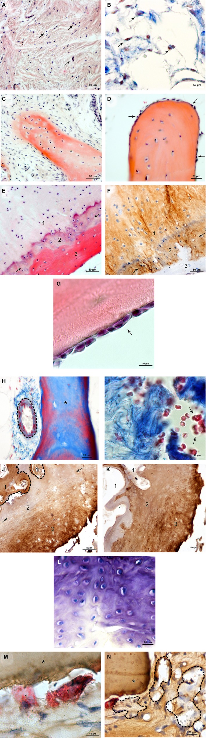
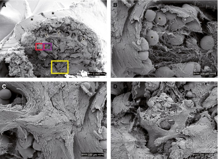
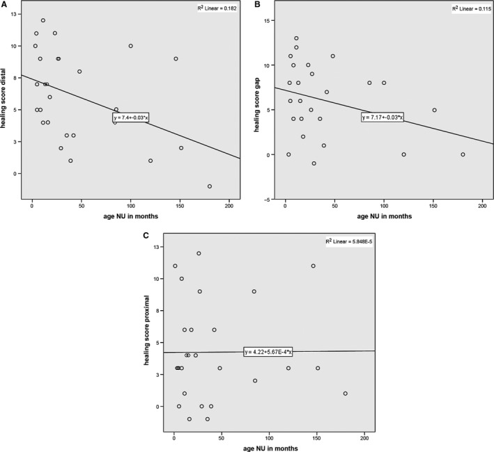
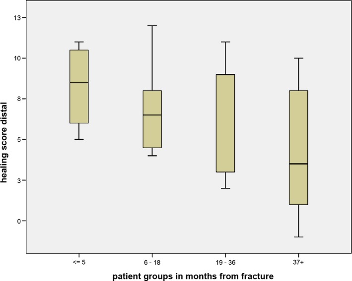
The scaphoid is the most frequently fractured carpal bone and prone to non-union due to mechanical and biological factors. Whereas the importance of stability is well documented, the evaluation of biological activity is mostly limited to the assessment of vascularity. The purpose of this study was to select histological and immunocytochemical parameters that could be used to assess healing potential after scaphoid fractures and to correlate these findings with time intervals after fracture for the three parts of the scaphoid (distal, gap and proximal). Samples were taken during operative intervention in 33 patients with delayed or non-union of the scaphoid. Haematoxylin and Eosin (HE), Azan, Toluidine, von Kossa and Tartrate-resistant acid phosphatase (TRAP) staining were used to characterise the samples histologically. We determined distribution of collagen 1 and 2 by immunocytochemistry, and scanning electron microscopy (SEM) was used to investigate the ultrastructure. To analyse the samples, parameters for biological healing status were defined and grouped according to healing capacity in parameters with high, partial and little biological activity. These findings allowed scoring of biological healing capacity, and the ensuing results were correlated with different time intervals after fracture. The results showed reduced healing capacity over time, but not all parts of the scaphoid were affected in the same way. For the distal fragment, regression analysis showed a statistically significant correlation between summarised healing activity scores and time from initial fracture (r = -0.427, P = 0.026) and decreasing healing activity for the gap region (r = -0.339, P = 0.090). In contrast, the analyses of the proximal parts for all patients did not show a correlation (r = 0.008, P = 0.969) or a decrease in healing capacity, with reduced healing capacity already at early stages. The histological and immunocytochemical characterisation of scaphoid non-unions (SNUs) and the scoring of healing parameters make it possible to analyse the healing capacity of SNUs at certain time points. This information is important as it can assist the surgeon in the selection of the most appropriate SNU treatment.

Keywords: bone healing capacity; histological characterisation; scaphoid fracture; scaphoid non-union; time dependent changes.


Figures

Figure 1
Figure 1
(A, B) Selected histological healing parameters in SNU samples: cell density, gap. (A) Connective tissue with densely packed cell nuclei (purple, arrows) and collagen fibres (pink) indicating highly active tissue; HE stain. (B) Less dense filled gap with fewer cell nuclei (red, arrows) and collagen fibres (blue) representing less active tissue; AZAN stain. Scale bars: 50 μm. (C, D) Selected histological healing parameters in SNU samples: trabecular spikes/thickening. (C) Trabecula formed by secreted extracellular matrix (red) and embedded osteocytes in typical spike formation; HE stain. (D) Osteoblasts (arrows) on the trabecular surface indicate trabecular thickening; HE stain. Scale bars: 50 μm (C), 20 μm (D). (E, F) Selected histological healing parameters in SNU samples: osteoid formation. (E) Cartilage calcification in the transition zone (2, arrows). Cartilage (1) transforms gradually into bone (3) with osteoid formation (red); HE stain. (F) Same sample with collagen 2 (brown) being present in the cartilage (1) and the transition zone (2), but not in bone (3); collagen 2. Scale bars: 50 μm. (G) Selected histological healing parameters in SNU samples: cell lines (osteoblasts). Lines of osteoblasts (purple, arrow) on the trabecular surface (red); HE stain. Scale bar:  10 μm. (H, I) Selected histological healing parameters in SNU samples: blood cells/vessels. Blood cells and blood vessels supply the tissue with nutrients, oxygen and precursor cells and are a prerequisite for healing. (H) Blood vessel (dashed line) next to a trabecula (asterisk); AZAN stain. (I) Blood cells (red, arrows) next to collagen fibres (blue); AZAN stain. Scale bars:  20 μm (H), 10 μm (I). (J,K) Selected histological healing parameters in SNU samples: collagen 1 (typical for bone) and collagen 2 (typical for cartilage), gap. The presence of both collagens was observed in some cases of SNU degradation. Predominance of collagen 2 next to the gap indicates formation of late SNU. (J) Collagen 1 (brown) is found in bone (dashed line, 1) and next to the gap (3) with unspecific cartilage stain in the middle portion (2); collagen 1. (K) The same sample shows collagen 2 (brown) in the middle portion (2) and next to the gap (3); collagen 2. Scale bars:  100 μm (J, K). (L) Selected histological healing parameters in SNU samples: cartilage formation, gap. Cartilage in the gap is a sign of encapsulation and SNU. Glycosaminoglycans (purple) and cell nuclei (blue) indicate the formation of cartilage; Toluidine stain. Scale bar: 20 μm. (M, N) Selected histological healing parameters in SNU samples: osteoclasts/foam deposits. High abundance of osteoclasts as well as foam deposits are a clear negative sign of bone healing capacity. (M) Osteoclasts (red, 1) next to a trabecula (asterisk); TRAP stain and collagen 1. (N) Foam deposits (dashed line) positive for collagen 1 (brown) next to an osteoclast (red, 1) and a trabecula (asterisk); TRAP stain and collagen 1. Scale bars: 10 μm (M), 20 μm (N).
Figure 2
Figure 2
SEM of the proximal pole of a long‐standing SNU. (A) Overview image showing an outer cartilage layer (asterisks), abundant subchondral fat deposits (arrows) and an overall degeneration of the bone tissue. Rectangular areas highlight the location of the magnified images. (B) Fat deposits (asterisks) within loosely organised fibres of connective tissue. (C) Undirected orientation and (D) spongy degeneration of the trabeculae. Scale bars: 1 mm (A), 100 μm (B,C), 200 μm (D).
Figure 3
Figure 3
Von Kossa stain of a whole unembedded sample shows (A) trabecular structure intact at the distal part of the scaphoid bone and (B) a loss of the trabecular structure at the proximal pole from the same patient.
Figure 4
Figure 4
(A) Scatter plot of healing activity score of the distal fragment vs. time from initial fracture with regression line (y = 7.4 − 0.03*x) and R 2 (R 2 = 0.182). (B) Scatter plot of healing activity score of the gap section vs. time from initial fracture with regression line (y = 7.17 − 0.03*x) and R 2 (R 2 = 0.115). (C) Scatter plot of healing activity score of the proximal part vs. time since initial fracture; no correlation or change over time is visible.
Figure 5
Figure 5
Box plot of healing activity score from the distal pole grouped by time since fracture. Central lines in each box denote the median, the lower/upper rims represent first/third quartiles and the whiskers extend to the minimum/maximum values.

References

    1. Bervian MR, Ribak S, Livani B (2015) Scaphoid fracture nonunion: correlation of radiographic imaging, proximal fragment histologic viability evaluation, and estimation of viability at surgery: diagnosis of scaphoid pseudarthrosis. Int Orthop 39, 67–72. - PubMed
    1. Beutel FK, Wilhelm K (1999) Relation of necrosis to outcome and site of fracture in scaphoid pseudarthrosis. Handchir Mikrochir Plast Chir 31, 207–211. - PubMed
    1. Blumer MJ, Hausott B, Schwarzer C, et al. (2012) Role of tartrate‐resistant acid phosphatase (TRAP) in long bone development. Mech Dev 129, 162–176. - PMC - PubMed
    1. Boyer MI, von Schroeder HP, Axelrod TS (1998) Scaphoid nonunion with avascular necrosis of the proximal pole. Treatment with a vascularized bone graft from the dorsum of the distal radius. J Hand Surg 23, 686–690. - PubMed
    1. Fernandez DL (1990) Anterior bone grafting and conventional lag screw fixation to treat scaphoid nonunions. J Hand Surg Am 15, 140–147. - PubMed