Identification of transcripts potentially involved in neural tube closure using RNA sequencing
- PMID: 29488319
- PMCID: PMC6616371
- DOI: 10.1002/dvg.23096
Identification of transcripts potentially involved in neural tube closure using RNA sequencing
Erratum in
-
Identification of transcripts potentially involved in neural tube closure using RNA sequencing.Genesis. 2019 Oct;57(10):e23332. doi: 10.1002/dvg.23332. Epub 2019 Aug 22. Genesis. 2019. PMID: 31603271 No abstract available.
Abstract
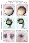
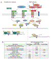
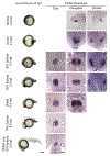
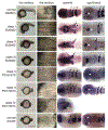
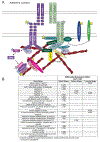
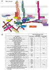
Anencephaly is a fatal human neural tube defect (NTD) in which the anterior neural tube remains open. Zebrafish embryos with reduced Nodal signaling display an open anterior neural tube phenotype that is analogous to anencephaly. Previous work from our laboratory suggests that Nodal signaling acts through induction of the head mesendoderm and mesoderm. Head mesendoderm/mesoderm then, through an unknown mechanism, promotes formation of the polarized neuroepithelium that is capable of undergoing the movements required for closure. We compared the transcriptome of embryos treated with a Nodal signaling inhibitor at sphere stage, which causes NTDs, to embryos treated at 30% epiboly, which does not cause NTDs. This screen identified over 3,000 transcripts with potential roles in anterior neurulation. Expression of several genes encoding components of tight and adherens junctions was significantly reduced, supporting the model that Nodal signaling regulates formation of the neuroepithelium. mRNAs involved in Wnt, FGF, and BMP signaling were also differentially expressed, suggesting these pathways might regulate anterior neurulation. In support of this, we found that pharmacological inhibition of FGF-receptor function causes an open anterior NTD as well as loss of mesodermal derivatives. This suggests that Nodal and FGF signaling both promote anterior neurulation through induction of head mesoderm.
Keywords: RNA sequencing; neural tube defects; neurulation; nodal; zebrafish.
© 2018 Wiley Periodicals, Inc.
Conflict of interest statement
The authors have no competing interests.
Figures




Similar articles
-
Temporal and spatial requirements for Nodal-induced anterior mesendoderm and mesoderm in anterior neurulation.Genesis. 2016 Jan;54(1):3-18. doi: 10.1002/dvg.22908. Epub 2016 Jan 12. Genesis. 2016. PMID: 26528772 Free PMC article.
-
An FGF3-BMP Signaling Axis Regulates Caudal Neural Tube Closure, Neural Crest Specification and Anterior-Posterior Axis Extension.PLoS Genet. 2016 May 4;12(5):e1006018. doi: 10.1371/journal.pgen.1006018. eCollection 2016 May. PLoS Genet. 2016. PMID: 27144312 Free PMC article.
-
Nodal signaling is required for closure of the anterior neural tube in zebrafish.BMC Dev Biol. 2007 Nov 8;7:126. doi: 10.1186/1471-213X-7-126. BMC Dev Biol. 2007. PMID: 17996054 Free PMC article.
-
G-protein-coupled receptor signaling and neural tube closure defects.Birth Defects Res. 2017 Jan 30;109(2):129-139. doi: 10.1002/bdra.23567. Birth Defects Res. 2017. PMID: 27731925 Free PMC article. Review.
-
Genetic backgrounds and modifier genes of NTD mouse models: An opportunity for greater understanding of the multifactorial etiology of neural tube defects.Birth Defects Res. 2017 Jan 30;109(2):140-152. doi: 10.1002/bdra.23554. Birth Defects Res. 2017. PMID: 27768235 Review.
Cited by
-
Grainyhead-like 2 as a double-edged sword in development and cancer.Am J Transl Res. 2020 Feb 15;12(2):310-331. eCollection 2020. Am J Transl Res. 2020. PMID: 32194886 Free PMC article. Review.
-
Neurotoxic effects in zebrafish embryos by valproic acid and nine of its analogues: the fish-mouse connection?Arch Toxicol. 2021 Feb;95(2):641-657. doi: 10.1007/s00204-020-02928-7. Epub 2020 Oct 27. Arch Toxicol. 2021. PMID: 33111190 Free PMC article.
-
Deciphering the enigma of the function of alpha-tocopherol as a vitamin.Free Radic Biol Med. 2024 Aug 20;221:64-74. doi: 10.1016/j.freeradbiomed.2024.05.028. Epub 2024 May 15. Free Radic Biol Med. 2024. PMID: 38754744 Free PMC article.
-
Nodal Enhances Perineural Invasion in Pancreatic Cancer by Promoting Tumor-Nerve Convergence.J Healthc Eng. 2022 Jan 27;2022:9658890. doi: 10.1155/2022/9658890. eCollection 2022. J Healthc Eng. 2022. PMID: 35126957 Free PMC article.
-
Understanding the Role of ATP Release through Connexins Hemichannels during Neurulation.Int J Mol Sci. 2023 Jan 21;24(3):2159. doi: 10.3390/ijms24032159. Int J Mol Sci. 2023. PMID: 36768481 Free PMC article.
References
-
- Alexander J, Stainier DY. 1999. A molecular pathway leading to endoderm formation in zebrafish. Curr Biol 9: 1147–1157. - PubMed
-
- Anders S, Huber W. 2012. Differential expression of RNA-Seq data at the gene level–the DESeq package. Heidelberg, Germany: European Molecular Biology Laboratory (EMBL).
MeSH terms
Substances
Grants and funding
LinkOut - more resources
Full Text Sources
Other Literature Sources
Medical
Molecular Biology Databases

