. 2018 May;17(5):6497-6505.
doi: 10.3892/mmr.2018.8642. Epub 2018 Feb 27.

Isoflurane reduces pain and inhibits apoptosis of myocardial cells through the phosphoinositide 3-kinase/protein kinase B signaling pathway in mice during cardiac surgery

Affiliations

Isoflurane reduces pain and inhibits apoptosis of myocardial cells through the phosphoinositide 3-kinase/protein kinase B signaling pathway in mice during cardiac surgery

Zhibing Pi et al. Mol Med Rep. 2018 May.

Retraction in

Abstract

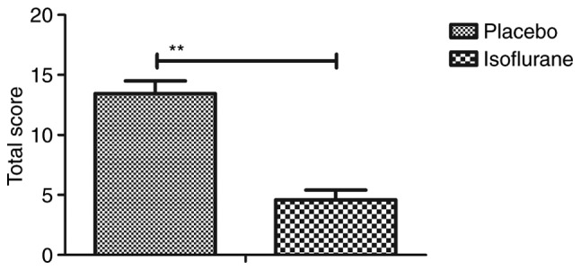
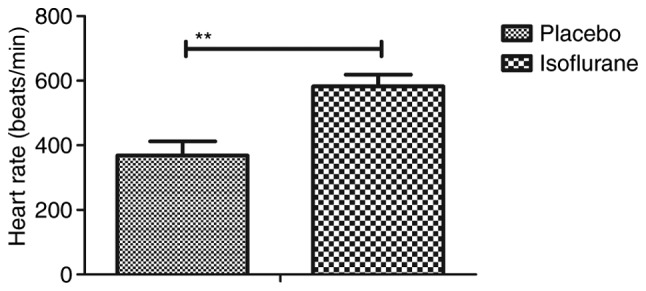
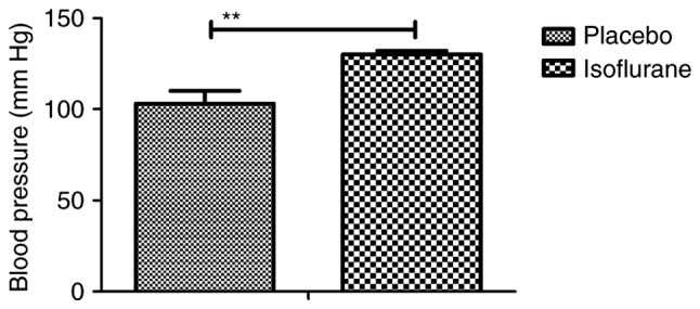
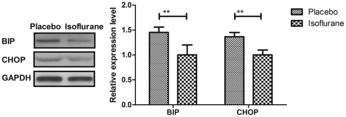
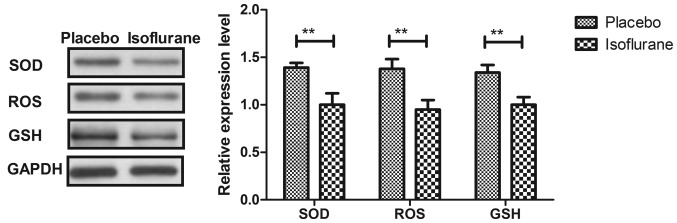
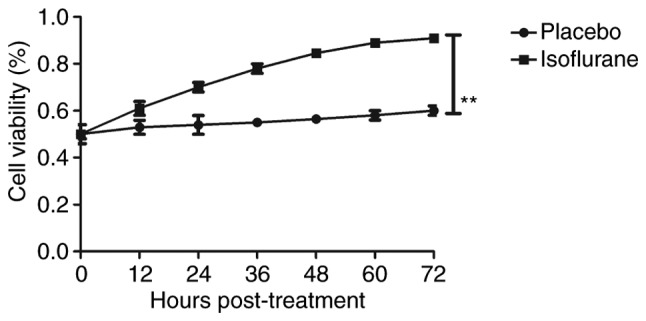
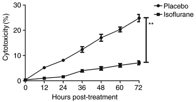
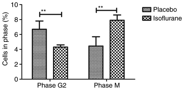
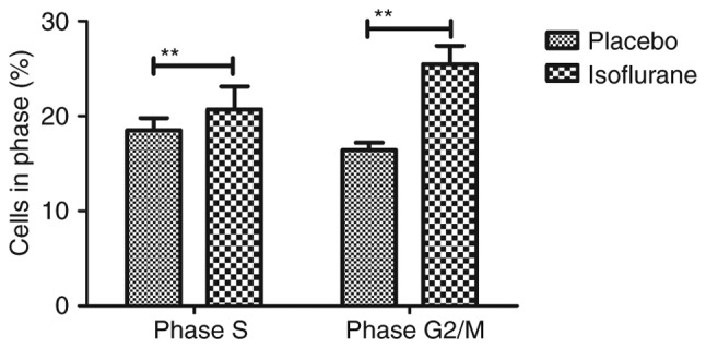
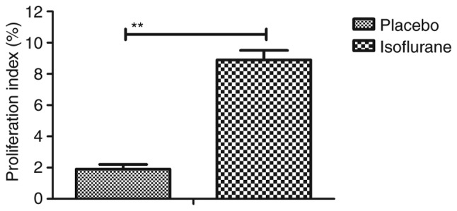
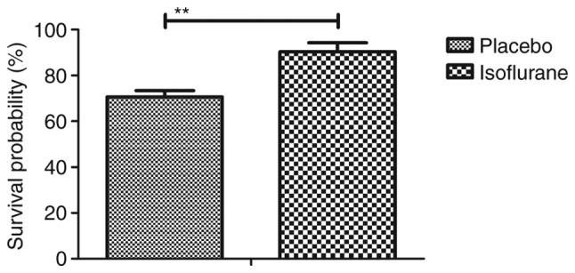
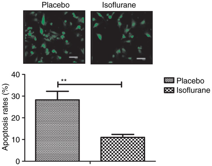
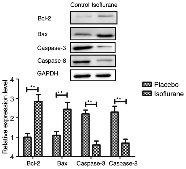
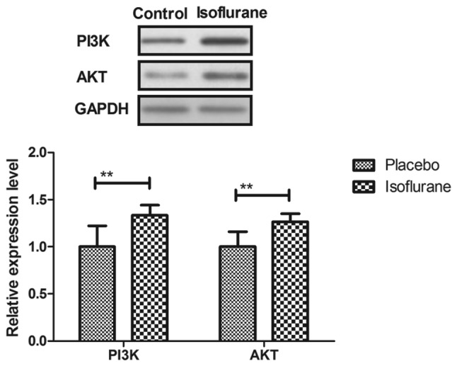
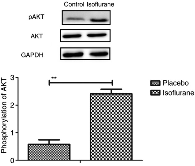
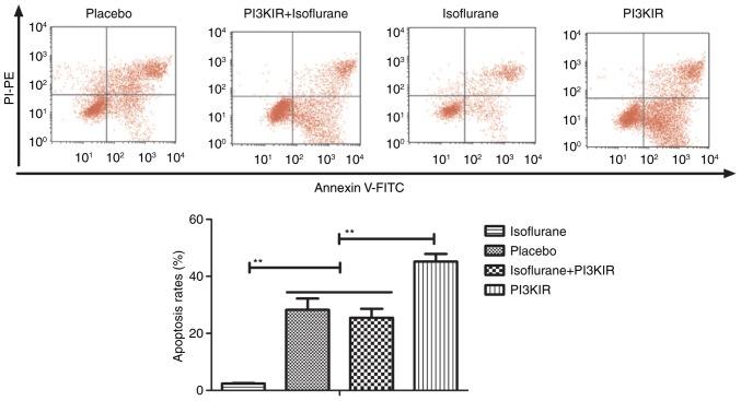
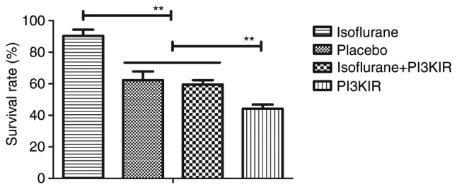
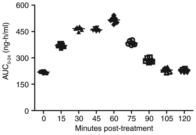
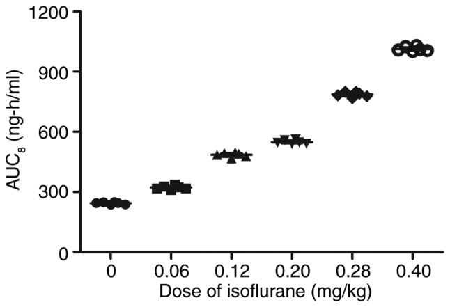
Heart bypass surgery is the most common treatment for myocardial ischemia. Clinical investigations have revealed that isoflurane anesthesia is efficient to alleviate pain during cardiac surgery, including heart bypass surgery. Previous studies have revealed the protective effects of isoflurane on myocardial cells of patients with myocardial ischemia during the perioperative period. The present study aimed to investigate the mechanism underlying the protective effects of isoflurane on myocardial cells in mice with myocardial ischemia. ELISA, flow cytometry, immunofluorescence and western blotting were used to analyze the effects of isoflurane anesthesia on myocardial cells. Briefly, myocardial cell apoptosis and viability, pain, phosphoinositide 3‑kinase/protein kinase B (PI3K/AKT) signaling pathway expression and the pharmacodynamics of isoflurane were studied in mice treated with isoflurane for heart bypass surgery. The results demonstrated that isoflurane anesthesia efficiently attenuated pain in mice during surgery. Viability and apoptosis of myocardial cells was also improved by isoflurane in vitro and in vivo. The PI3K/AKT pathway was upregulated in myocardial cells on day 3 post‑operation. Mechanistically, isoflurane promoted PI3K/AKT activation, upregulated B‑cell lymphoma 2 (Bcl‑2)‑associated X protein and Bcl‑2 expression levels, and reduced the expression levels of caspase‑3 and caspase‑8 in myocardial cells. In conclusion, the findings indicated that isoflurane is beneficial for pain attenuation and inhibits apoptosis of myocardial cells via the PI3K/AKT signaling pathway in mice during cardiac surgery.

Keywords: isoflurane; pain; myocardial ischemia; myocardial cells; apoptosis; phosphoinositide 3-kinase/protein kinase B.


Figures

Figure 1.
Figure 1.
General appearance parameters total score for C57BL/6 mice (n=10/group) with myocardial ischemia following heart bypass surgery. Data are presented as the mean + standard error of the mean of three independent experiments. **P<0.01 vs. the placebo group.
Figure 2.
Figure 2.
Heart rate of C57BL/6 mice (n=10/group) with myocardial ischemia following heart bypass surgery. Data are presented as the mean + standard error of the mean of three independent experiments. **P<0.01 vs. the placebo group.
Figure 3.
Figure 3.
Arterial blood pressure of C57BL/6 mice (n=10/group) with myocardial ischemia following heart bypass surgery. Data are presented as the mean + standard error of the mean of three independent experiments. **P<0.01 vs. the placebo group.
Figure 4.
Figure 4.
Protein expression levels of BIP and CHOP in myocardial cells obtained from mice with myocardial ischemia following heart bypass surgery. Data are presented as the mean + standard error of the mean of three independent experiments. **P<0.01 vs. the placebo group. BIP, binding immunoglobulin protein; CHOP, CCAAT-enhancer-binding protein homologous protein.
Figure 5.
Figure 5.
Protein expression levels of, SOD, ROS and GSH in myocardial cells from mice with myocardial ischemia following heart bypass surgery. Data are presented as the mean + standard error of the mean of three independent experiments. **P<0.01 vs. the placebo group. GSH, glutathione; ROS, proto-oncogene tyrosine-protein kinase ROS; SOD, superoxide dismutase.
Figure 6.
Figure 6.
Viability of myocardial cells from mice with myocardial ischemia following heart bypass surgery and isoflurane treatment. Data are presented as the mean + standard error of the mean of three independent experiments. **P<0.01 vs. the placebo group.
Figure 7.
Figure 7.
Cytotoxicity of isoflurane on myocardial cells from mice with myocardial ischemia following heart bypass surgery. Data are presented as the mean + standard error of the mean of three independent experiments. **P<0.01 vs. the placebo group.
Figure 8.
Figure 8.
Effects of isoflurane on the number of myocardial cells in G2 and M phases. Cells were isolated from mice with myocardial ischemia following heart bypass surgery and isoflurane treatment. Data are presented as the mean + standard error of the mean of three independent experiments. **P<0.01 vs. the placebo group.
Figure 9.
Figure 9.
Effects of isoflurane on the number of myocardial cells in S and G2/M phases. Cells were isolated from mice with myocardial ischemia following heart bypass surgery and isoflurane treatment. Data are presented as the mean + standard error of the mean of three independent experiments. **P<0.01 vs. the placebo group.
Figure 10.
Figure 10.
Effects of isoflurane on the proliferation of myocardial cells from mice with myocardial ischemia following heart bypass surgery. Data are presented as the mean + standard error of the mean of three independent experiments. **P<0.01 vs. the placebo group.
Figure 11.
Figure 11.
Survival of myocardial cells isolated from mice with myocardial ischemia following heart bypass surgery and isoflurane treatment. Data are presented as the mean + standard error of the mean of three independent experiments. **P<0.01 vs. the placebo group.
Figure 12.
Figure 12.
Apoptotic rate of myocardial cells, as determined by terminal deoxynucleotidyl-transferase-mediated dUTP nick-end labeling assay. Data are presented as the mean + standard error of the mean of three independent experiments. **P<0.01 vs. the placebo group.
Figure 13.
Figure 13.
Expression levels of Bax and Bcl-2, caspase-3 and caspase-8 in myocardial cells isolated from mice with myocardial ischemia following heart bypass surgery and isoflurane treatment. Data are presented as the mean + standard error of the mean of three independent experiments. **P<0.01 vs. the placebo group. Bax, Bcl-2-associated X protein; Bcl-2, B-cell lymphoma 2.
Figure 14.
Figure 14.
Expression levels of PI3K and AKT in myocardial cells isolated from mice with myocardial ischemia following heart bypass surgery and isoflurane treatment. Data are presented as the mean + standard error of the mean of three independent experiments. **P<0.01 vs. the placebo group. AKT, protein kinase B; PI3K, phosphoinositide 3-kinase.
Figure 15.
Figure 15.
Phosphorylation of AKT in myocardial cells isolated from mice with myocardial ischemia following heart bypass surgery and isoflurane treatment. Data are presented as the mean + standard error of the mean of three independent experiments. **P<0.01 the placebo group. AKT, protein kinase B; pAKT, phosphorylated-AKT.
Figure 16.
Figure 16.
PI3KIR abolishes isoflurane-induced inhibition of apoptosis of myocardial cells obtained from experimental mice. Data are presented as the mean + standard error of the mean of three independent experiments.**P<0.01. FITC, fluorescein isothiocyanate; PI3KIR, phosphoinositide 3-kinase inhibitor; PI-PE, propidium iodide-phycoerythrin.
Figure 17.
Figure 17.
PI3KIR reduces isoflurane-stimulated myocardial cell survival. Data are presented as the mean + standard error of the mean of three independent experiments. **P<0.01. PI3KIR, phosphoinositide 3-kinase inhibitor.
Figure 18.
Figure 18.
Serum concentration of isoflurane in mice with myocardial ischemia that underwent heart bypass surgery following isoflurane treatment. Data are presented as the mean + standard error of the mean of three independent experiments. AUC, area under the curve.
Figure 19.
Figure 19.
Cmax concentrations of isoflurane (0–0.40 mg/kg) in mice with myocardial ischemia that underwent heart bypass surgery following isoflurane treatment. Data are presented as the mean + standard error of the mean of three independent experiments. AUC, area under the curve.

References

    1. Yang CJ, Yang J, Fan ZX. Activating transcription factor 3-an endogenous inhibitor of myocardial ischemia-reperfusion injury (Review) Mol Med Rep. 2016;13:9–12. doi: 10.3892/mmr.2015.4529. - DOI - PubMed
    1. Yang Y, Sun Y, Yi W, Li Y, Fan C, Xin Z, Jiang S, Di S, Qu Y, Reiter RJ, Yi D. A review of melatonin as a suitable antioxidant against myocardial ischemia-reperfusion injury and clinical heart diseases. J Pineal Res. 2014;57:357–366. doi: 10.1111/jpi.12175. - DOI - PubMed
    1. Wang N, Min X, Li D, He P, Zhao L. Geranylgeranylacetone protects against myocardial ischemia and reperfusion injury by inhibiting high-mobility group box 1 protein in rats. Mol Med Rep. 2012;5:521–524. doi: 10.3892/mmr.2012.745. - DOI - PubMed
    1. Yao HC, Zhou M, Zhou YH, Wang LH, Zhang DY, Han QF, Liu T, Wu L, Tian KL, Zhang M. Intravenous high mobility group box 1 upregulates the expression of HIF-1α in the myocardium via a protein kinase B-dependent pathway in rats following acute myocardial ischemia. Mol Med Rep. 2016;13:1211–1219. doi: 10.3892/mmr.2015.4648. - DOI - PMC - PubMed
    1. He F, Xu BL, Chen C, Jia HJ, Wu JX, Wang XC, Sheng JL, Huang L, Cheng J. Methylophiopogonanone A suppresses ischemia/reperfusion-induced myocardial apoptosis in mice via activating PI3K/Akt/eNOS signaling pathway. Acta Pharmacol Sin. 2016;37:763–771. doi: 10.1038/aps.2016.14. - DOI - PMC - PubMed

Publication types

Substances