Review
. 2018 Apr 1;98(2):813-880.
doi: 10.1152/physrev.00011.2017.

Neuronal Cell Death

Affiliations
Review

Neuronal Cell Death

Michael Fricker et al. Physiol Rev. .

Abstract

Neuronal cell death occurs extensively during development and pathology, where it is especially important because of the limited capacity of adult neurons to proliferate or be replaced. The concept of cell death used to be simple as there were just two or three types, so we just had to work out which type was involved in our particular pathology and then block it. However, we now know that there are at least a dozen ways for neurons to die, that blocking a particular mechanism of cell death may not prevent the cell from dying, and that non-neuronal cells also contribute to neuronal death. We review here the mechanisms of neuronal death by intrinsic and extrinsic apoptosis, oncosis, necroptosis, parthanatos, ferroptosis, sarmoptosis, autophagic cell death, autosis, autolysis, paraptosis, pyroptosis, phagoptosis, and mitochondrial permeability transition. We next explore the mechanisms of neuronal death during development, and those induced by axotomy, aberrant cell-cycle reentry, glutamate (excitoxicity and oxytosis), loss of connected neurons, aggregated proteins and the unfolded protein response, oxidants, inflammation, and microglia. We then reassess which forms of cell death occur in stroke and Alzheimer's disease, two of the most important pathologies involving neuronal cell death. We also discuss why it has been so difficult to pinpoint the type of neuronal death involved, if and why the mechanism of neuronal death matters, the molecular overlap and interplay between death subroutines, and the therapeutic implications of these multiple overlapping forms of neuronal death.


Figures

FIGURE 1.
FIGURE 1.
Overview of apoptosis. The internal (mitochondrial) pathway of apoptosis is triggered within the cell, causing expression or activation of BH3-only proteins that activate Bax (and/or Bak in some cells) to form pores in the outer mitochondrial membrane, releasing cytochrome c to bind APAF-1, activating caspase-9 to cleave and activate downstream caspases, which degrades cellular proteins. The external (death receptor) pathway starts outside the cell with death ligands activating death receptors to activate caspase-8, which either cleaves downstream caspases or cleaves and activates the BH3-only protein Bid. Anti-apoptotic proteins, such as Bcl-2, hold inactive Bax or BH3-ony proteins.
FIGURE 2.
FIGURE 2.
Bax signaling at the mitochondria. BH3-only proteins activate Bax to oligomerize and form pores in the outer mitochondrial membrane, causing cytochrome c release and inhibition of complex II, inhibition of respiration and ROS production, activating the protease OMA-1 to remodel the inner mitochondrial membrane, which enables greater cytochrome c release, which triggers caspase activation and apoptosis.
FIGURE 3.
FIGURE 3.
Oncosis. Ischemia, mitochondrial dysfunction, and/or excessive ATP consumption cause cellular ATP depletion, resulting in 1) failure of sodium pump resulting in cell swelling to rupture, and 2) failure of calcium pumps resulting in calcium activation of proteases and phospholipases that degrade the cell.
FIGURE 4.
FIGURE 4.
Necroptosis. Activation of death receptors or Toll-like receptors activates NF-κB-mediated inflammation via ubiquinated RIPK1, but deubiquinated RIPK1 can form a complex with RIPK3 that can induce necrosis if and only if caspase-8 is inhibited, preventing cleavage of RIPK1. RIPK1 phosphorylates RIPK3, which phosphorylates MLKL1, which permeabilizes membranes.
FIGURE 5.
FIGURE 5.
Parthanatos: poly ADP-ribose polymerase 1 (PARP-1)-mediated cell death.
FIGURE 6.
FIGURE 6.
Ferroptosis. Ferroptosis results from excessive peroxidation of membrane lipids, promoted by reduced iron (Fe2+) and inhibited by glutathione peroxidase 4, which depends on glutathione, which in turn depends on cystine supplied by the cystine/glutamate exchanger (Xc). Red text indicates inducers of ferroptosis, and green text indicates inhibitors of ferroptosis.
FIGURE 7.
FIGURE 7.
Cell death by mitochondrial permeability transition. Calcium and ROS trigger formation of the permeability transition pore (mPTP) in the inner mitochondrial membrane, consisting probably of the adenine nucleotide carrier (ANT) and cyclophilin D (CypD), or possibly the phosphate carrier (PiC) or ATP synthase, and regulated by VDAC and hexokinase on the outer membrane. mPTP causes ATP depletion and thus necrosis, but also may cause cytochrome c release to trigger apoptosis if sufficient ATP is still present.
FIGURE 8.
FIGURE 8.
Lysosomal cell death. Multiple stimuli can cause lysosomal membrane permeabilization (LMP), releasing cathepsin proteases that induce cell death by multiple routes. LMP may also cause the release of calcium, which activates calpain, as well as DNase II and other hydrolases (e.g., lipases).
FIGURE 9.
FIGURE 9.
Pyroptosis. Inflammation causes expression and activation of the inflammasome, causing activation of caspase-1, which cleaves 1) pro-IL-1β to IL-1β, a key inflammatory cytokine, and 2) gasdermin D (and/or DFNA5) to an NH2-terminal fragment that oligomerizes into pores.
FIGURE 10.
FIGURE 10.
Autophagy and autophagic cell death. Autophagy normally promotes survival during starvation or growth factor withdrawal, but, if excessive, can cause autophagic cell death, characterized by the accumulation of autophagic vacuoles. Note the cross talk between autophagy and apoptosis as Beclin-1 is held via its BH3 motif in an inactive state by binding to anti-apoptotic Bcl2 family members or to Bim when it is tethered on microtubules.
FIGURE 11.
FIGURE 11.
Phagoptosis is cell death resulting from phagocytosis of the cell. Neuronal stress can result in exposure of ‟eat me” signals such as phosphatidylserine, which bind opsonins such as MFG-E8 or GAS6 that engage phagocytic receptors VNR (vitronectin receptor) and MERTK, respectively. Complement factors C1 and C3 binding to neurons can induce uptake via CR3. Phagoptosis is increased in inflammatory conditions that stress neurons and increase phagocytosis.
FIGURE 12.
FIGURE 12.
Neuronal death induced by axotomy.
FIGURE 13.
FIGURE 13.
Excitotoxicity. Glutamate induces neuronal death by multiple mechanisms. Autophagic cell death may also contribute. Outcome is dependent on neuronal type, stimulus strength and duration, preconditioning, age, gender, etc.
FIGURE 14.
FIGURE 14.
Neuronal death induced by protein aggregates. Protein aggregates can trigger the unfolded protein response (UPR) or form membrane pores that elevate calcium and ROS levels, which can trigger cell death by multiple mechanisms. Protein aggregates can also clog protein import into organelles, induce stress granule formation, interfere with RNA and heat shock proteins, and impair the proteasome/autophagy, but how these connect to specific cell death pathways is not clear.
FIGURE 15.
FIGURE 15.
Brain ischemia. Brain ischemia induces a necrotic core, delayed neuronal death in the penumbra (where hypoxia is not so deep), and secondary neuronal loss in connected areas of the brain.
FIGURE 16.
FIGURE 16.
Forms of ischemia-induced neuronal death in penumbra.
FIGURE 17.
FIGURE 17.
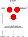
Alzheimer’s disease pathology and links to neuronal death.
FIGURE 18.
FIGURE 18.
Summary of some mechanisms of neuronal death.
FIGURE 19.
FIGURE 19.
The pores of death.

References

    1. Adalbert R, Nógrádi A, Szabó A, Coleman MP. The slow Wallerian degeneration gene in vivo protects motor axons but not their cell bodies after avulsion and neonatal axotomy. Eur J Neurosci 24: 2163–2168, 2006. doi:10.1111/j.1460-9568.2006.05103.x. - DOI - PubMed
    1. Adamczak SE, de Rivero Vaccari JP, Dale G, Brand FJ III, Nonner D, Bullock MR, Dahl GP, Dietrich WD, Keane RW. Pyroptotic neuronal cell death mediated by the AIM2 inflammasome. J Cereb Blood Flow Metab 34: 621–629, 2014. doi:10.1038/jcbfm.2013.236. - DOI - PMC - PubMed
    1. Ahmad M, Dar NJ, Bhat ZS, Hussain A, Shah A, Liu H, Graham SH. Inflammation in ischemic stroke: mechanisms, consequences and possible drug targets. CNS Neurol Disord Drug Targets 13: 1378–1396, 2014. doi:10.2174/1871527313666141023094720. - DOI - PubMed
    1. Airaksinen MS, Titievsky A, Saarma M. GDNF family neurotrophic factor signaling: four masters, one servant? Mol Cell Neurosci 13: 313–325, 1999. doi:10.1006/mcne.1999.0754. - DOI - PubMed
    1. Aits S, Jäättelä M. Lysosomal cell death at a glance. J Cell Sci 126: 1905–1912, 2013. doi:10.1242/jcs.091181. - DOI - PubMed

Publication types

LinkOut - more resources