. 2018 Mar 8;555(7695):265-268.
doi: 10.1038/nature25787. Epub 2018 Feb 28.

The mechanism of eukaryotic CMG helicase activation

Affiliations

The mechanism of eukaryotic CMG helicase activation

Max E Douglas et al. Nature. .

Abstract

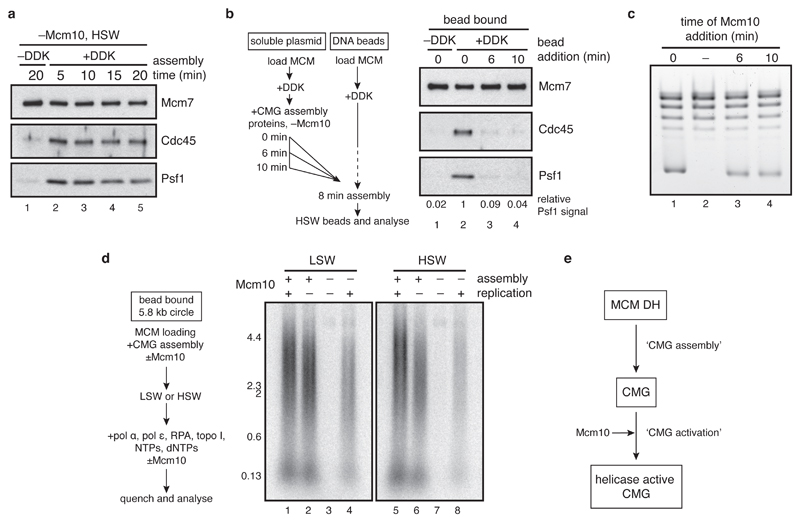
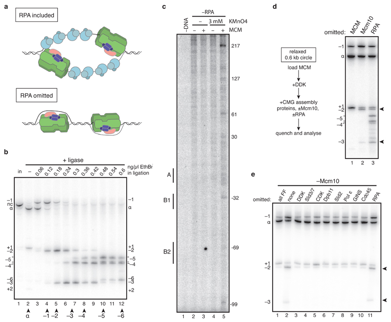
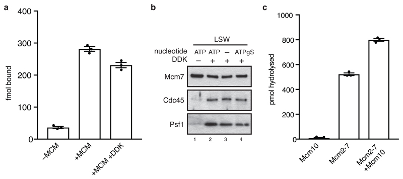
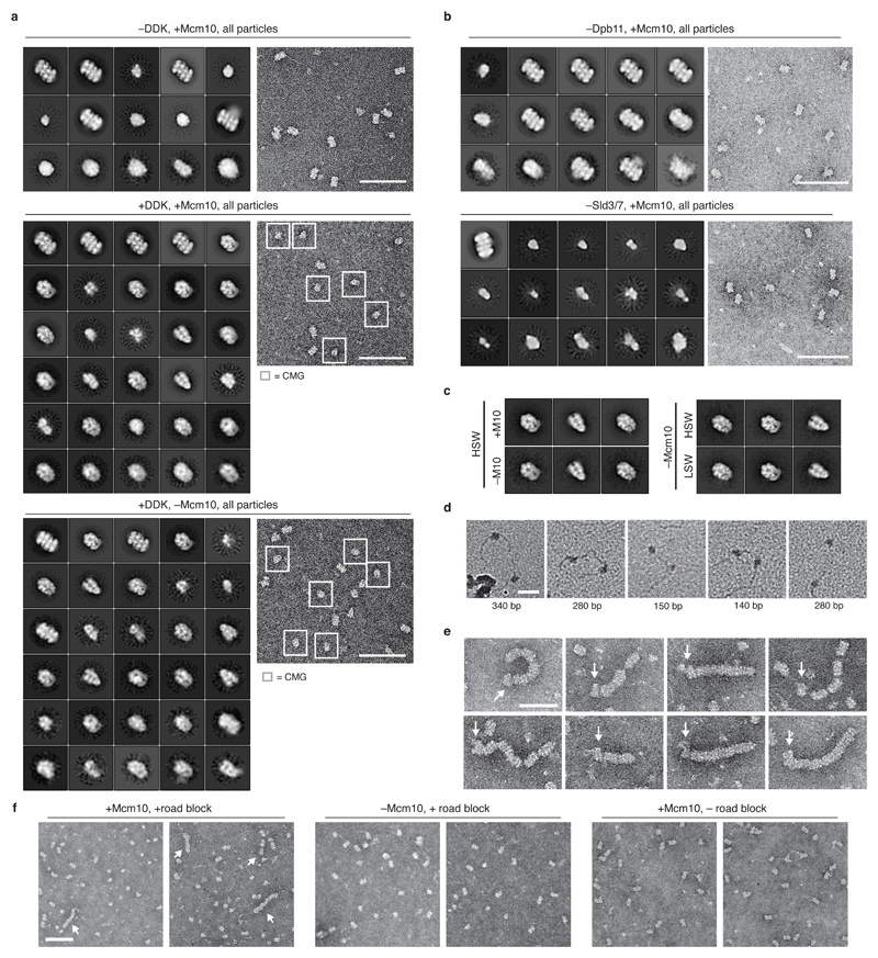
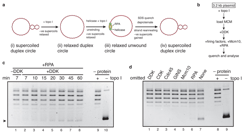
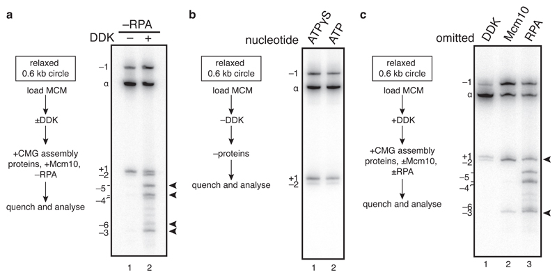
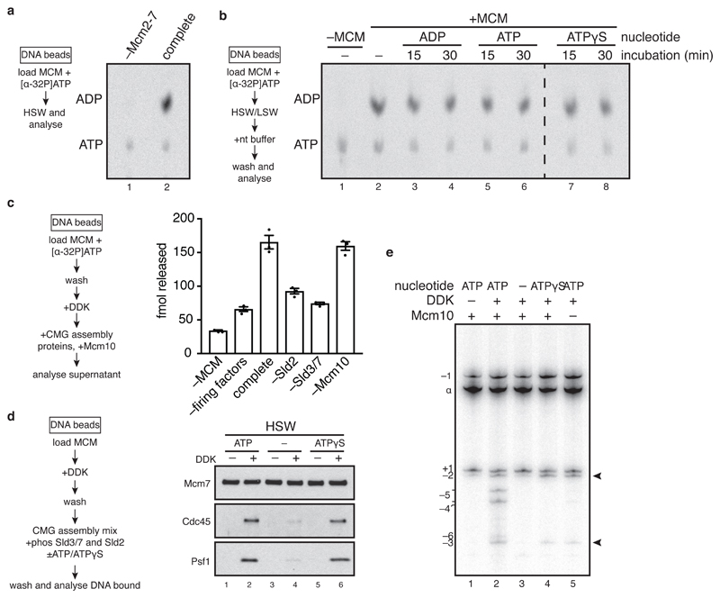
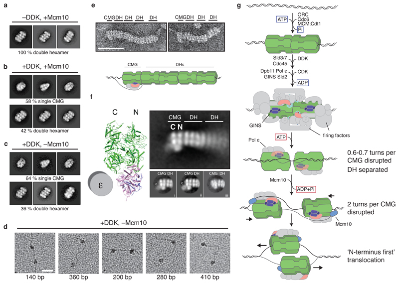
The initiation of eukaryotic DNA replication occurs in two discrete stages: first, the minichromosome maintenance (MCM) complex assembles as a head-to-head double hexamer that encircles duplex replication origin DNA during G1 phase; then, 'firing factors' convert each double hexamer into two active Cdc45-MCM-GINS helicases (CMG) during S phase. This second stage requires separation of the two origin DNA strands and remodelling of the double hexamer so that each MCM hexamer encircles a single DNA strand. Here we show that the MCM complex, which hydrolyses ATP during double-hexamer formation, remains stably bound to ADP in the double hexamer. Firing factors trigger ADP release, and subsequent ATP binding promotes stable CMG assembly. CMG assembly is accompanied by initial DNA untwisting and separation of the double hexamer into two discrete but inactive CMG helicases. Mcm10, together with ATP hydrolysis, then triggers further DNA untwisting and helicase activation. After activation, the two CMG helicases translocate in an 'N terminus-first' direction, and in doing so pass each other within the origin; this requires that each helicase is bound entirely to single-stranded DNA. Our experiments elucidate the mechanism of eukaryotic replicative helicase activation, which we propose provides a fail-safe mechanism for bidirectional replisome establishment.


Conflict of interest statement

The authors declare no competing financial interests.

Figures

Extended Data Figure 1
CMG assembly and activation are separable steps. a, to determine when CMG assembly saturates in our reactions, CMG assembly was carried out on bead-immobilised ARS1 DNA and washed with high salt wash buffer (HSW, buffer A +KCl) at the times indicates. The data show that no new CMG assembly takes place after 5 minutes. b, to confirm this, MCMs were loaded in parallel onto a bead-immobilised ARS1 DNA fragment and a soluble ARS1 plasmid, and phosphorylated with DDK. A firing factor mix that was complete except for Mcm10 was added to the soluble reaction only, which was then added to the bead-immobilised MCMs at the times indicated after firing factor addition to the soluble reaction. After 8 min, beads were washed with high salt (buffer A + KCl) and bound proteins analysed by immunoblotting. Psf1 signal relative to lane 2 is indicated. The experiment confirms that no CMG assembly can take place 5 minutes after firing factors have been added. c, to test whether Mcm10 can trigger DNA unwinding even after CMG assembly was finished, reactions were set up as in Figure 1d, except Mcm10 was omitted until the times indicated after firing factor addition. Mcm10 triggered robust unwinding, even when added more than 5 minutes after firing factors. Mcm10 can therefore activate preassembled CMG for DNA unwinding. d, to test whether Mcm10 can activate preassembled CMG for replication, CMG was assembled on an immobilised ARS1 plasmid ±Mcm10. Beads were washed with low (Buffer A + 0.25 M K-glutamate) or high (Buffer A + KCl) salt buffer, and replication proteins ±Mcm10 and cofactors including radiolabelled dCTP were added. Mcm10 enabled DNA replication even when CMG had been washed to remove excess firing factors. e, schematic outlining the CMG assembly and CMG activation steps described here.
Extended Data Figure 2
a, models of DNA unwinding ±RPA. b, to define the relative position of different topoisomers of radiolabelled 616 bp DNA circles containing ARS1 (used to analyse small changes in DNA supercoiling in the unwinding assay), nicked circles (nc, lane 1) were ligated closed under the ethidium bromide concentrations indicated. The supercoiling states of different bands of covalently closed DNA were determined relative to the ground state (α) by tracking the order in which bands peaked as ethidium bromide concentration increased and DNA was increasingly negatively supercoiled (see methods for further details). Two bands peak at the same position for α-5 and likely represent alternative configurations of the α-5 topoisomer. c, primer extension reactions reading the T-rich strand of the ARS-consensus sequence (ACS) of ARS1 were carried out on 616 bp ARS1 DNA treated with potassium permanganate as indicated after CMG assembly in the absence of RPA. Reactions were separated on 5 % sequencing gels, dried and analysed by autoradiography. Base pair numbering is relative to the 5’ end of the T-rich strand of the ACS. d, as in Figure 2c. Lane 1 shows that MCM loading is required for all shifts in topoisomer distribution. Compared with other control samples, such as –DDK, topoisomer distribution is subtly different without MCM; this is not due to loading, which, as shown in Figure 2b, does not affect topoisomer distribution. e, as in Figure 2a, except Mcm10 was omitted from all reactions. No proteins except topoisomerase were added to the reaction in lane 1 after MCM loading. No detectable change in supercoiling relative to when no firing factors were added (lane 1) was observed when each individual firing factor was omitted, suggesting DNA untwisting without Mcm10 takes place during CMG assembly.
Extended Data Figure 3
a, DHs assembled on bead-immobilised DNA using [α32P]ATP were treated with DDK as indicated and analysed by scintillation counting. Error bars represent standard error of the mean. b, immunoblots of CMG assembly reactions carried out as in Figure 3d and washed with low salt wash buffer (Buffer A + 0.25 M K-glutamate). c, ATPase assays using [α32P]ATP, single MCM hexamers and Mcm10 as indicated were quantified after thin layer chromatography. Error bars represent standard error of the mean.
Extended Data Figure 4
a, example micrographs and complete sets of reference free class averages of the helicase activation reactions indicated, washed with high salt wash buffer (buffer A + KCl). –DDK, +Mcm10, 7410 of 23092 total particles were DH. +DDK, +Mcm10, of 43320 total particles, 14668 and 10492 were CMG and DH respectively. +DDK, –Mcm10, of 12920 total particles, 3984 and 2226 were CMG and DH respectively. Classes are positioned with respect to the abundance of source particles, with the most abundant class in the top left-hand corner, and abundance decreasing from left to right and top to bottom. b, as in a. with representative source micrographs. 5032/6815 and 2049/20904 particles were DH when Dpb11 or Sld3/7 were omitted respectively. Scale bar represents 100 nm. c, comparison of CMG formed under the conditions indicated. HSW is buffer A + KCl, LSW is buffer A + 0.25 M K-glutamate. d, as in Figure 4d. e, as in Figure 4e. Arrows mark position of CMG. f, representative crops from micrographs imaging the samples indicated. MCM trains are marked with arrows. Trains were not observed when Mcm10 or the protein road block was omitted. Scale bar is 100 nm.
Figure 1
Figure 1. Analysis of replicative helicase activation with a DNA unwinding assay.
a, b, outline of the assay. c, time course of unwinding. Purified DNA products were separated on a native agarose gel and stained with ethidium bromide. No loading or firing factors were added to ‘–protein’ reactions. d, as in c, with proteins omitted as indicated. Reactions were quenched after 40 min.
Figure 2
Figure 2. Origin unwinding takes place in two steps.
a, active CMG was assembled on a radiolabelled 616 bp ARS1 circle without RPA for 40 min and products separated on a native bis-polyacrylamide gel. b, as in a, except all firing factors were omitted. MCM loading does not occur in ATPγS. c, as in a, with proteins omitted as indicated.
Figure 3
Figure 3. CMG assembly and activation are coupled to ATP binding and hydrolysis.
a, MCM loading reactions containing [α32P]ATP were washed with high salt (buffer A + NaCl) and analysed by thin layer chromatography. b, as in a. except washed reactions were incubated with nucleotide as indicated and washed again before analysis. c, DHs assembled on bead-immobilised DNA with [α32P]ATP were used in CMG assembly reactions, the supernatants from which were analysed by scintillation counting. Error bars show standard error of the mean. d, immunoblots of CMG assembly reactions carried out as in Extended Data Figure 1a, except CDK was omitted, Sic1 was added, Sld2 and Sld3/7 prephosphorylated with CDK were used, and the nucleotide indicated was added. Reactions were quenched 15 min after firing factor addition. e, as in d. except reactions were carried out on soluble ARS1 circles and analysed as in Figure 2a. ATP was removed after DDK phosphorylation using a spin column.
Figure 4
Figure 4. Structural characterisation of replicative helicase activation.
a-c, representative reference free class averages of helicase activation reactions washed with high salt (buffer A + KCl). DH and CMG classes are shown. All particle classes are presented in Extended Data Figure 4a. d, helicase activation reactions lacking Mcm10 were washed with high salt and positively stained to visualise DNA. Examples of two CMG-sized particles co-localised with a single DNA fragment are shown. The approximate base pair distance between particles is indicated. Scale bar represents 50 nm. e, examples of CMGs neighbouring DH ‘trains’ in high salt washed reactions on roadblocked DNA. Scale bar is 50 nm. f, annotated reference free class average from 469 train ends. CMG structure (from PDB file 3jc5) is included for reference. 2D classification of train tip particles into several classes (i-iii) reveals doughnut-shaped polymerase ε density on a subset of CMGs (i and ii). g, model of eukaryotic replicative helicase assembly and activation.

References

    1. Bell SP, Labib K. Chromosome Duplication in Saccharomyces cerevisiae. Genetics. 2016;203:1027–1067. - PMC - PubMed
    1. Coster G, Frigola J, Beuron F, Morris EP, Diffley JFX. Origin Licensing Requires ATP Binding and Hydrolysis by the MCM Replicative Helicase. Mol Cell. 2014;55:666–677. - PMC - PubMed
    1. Kang S, Warner MD, Bell SP. Multiple functions for Mcm2-7 ATPase motifs during replication initiation. Mol Cell. 2014;55:655–665. - PMC - PubMed
    1. Ilves I, Petojevic T, Pesavento JJ, Botchan MR. Activation of the MCM2-7 helicase by association with Cdc45 and GINS proteins. Mol Cell. 2010;37:247–258. - PMC - PubMed
    1. Georgescu RE, et al. Mechanism of asymmetric polymerase assembly at the eukaryotic replication fork. Nat Struct Mol Biol. 2014;21:664–670. - PMC - PubMed

Publication types

MeSH terms