. 2018 Mar 15;555(7696):382-386.
doi: 10.1038/nature25974. Epub 2018 Feb 28.

Recognition of DHN-melanin by a C-type lectin receptor is required for immunity to Aspergillus

Affiliations

Recognition of DHN-melanin by a C-type lectin receptor is required for immunity to Aspergillus

Mark H T Stappers et al. Nature. .

Abstract

Resistance to infection is critically dependent on the ability of pattern recognition receptors to recognize microbial invasion and induce protective immune responses. One such family of receptors are the C-type lectins, which are central to antifungal immunity. These receptors activate key effector mechanisms upon recognition of conserved fungal cell-wall carbohydrates. However, several other immunologically active fungal ligands have been described; these include melanin, for which the mechanism of recognition is hitherto undefined. Here we identify a C-type lectin receptor, melanin-sensing C-type lectin receptor (MelLec), that has an essential role in antifungal immunity through recognition of the naphthalene-diol unit of 1,8-dihydroxynaphthalene (DHN)-melanin. MelLec recognizes melanin in conidial spores of Aspergillus fumigatus as well as in other DHN-melanized fungi. MelLec is ubiquitously expressed by CD31+ endothelial cells in mice, and is also expressed by a sub-population of these cells that co-express epithelial cell adhesion molecule and are detected only in the lung and the liver. In mouse models, MelLec was required for protection against disseminated infection with A. fumigatus. In humans, MelLec is also expressed by myeloid cells, and we identified a single nucleotide polymorphism of this receptor that negatively affected myeloid inflammatory responses and significantly increased the susceptibility of stem-cell transplant recipients to disseminated Aspergillus infections. MelLec therefore recognizes an immunologically active component commonly found on fungi and has an essential role in protective antifungal immunity in both mice and humans.


Conflict of interest statement

The authors declare no competing financial interests.

Figures
Extended data Figure 1
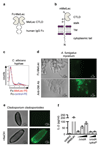
Extended data Figure 1. MelLec recognises ligands on selected fungi and fungal morphotypes.
Cartoon representation of the structure of Fc-MelLec (a) and the full length receptor (b). Lollipop structures represent predicted glycosylation sites. c, Fc-MelLec or Fc-CLEC12b (Fc-control) staining of C. albicans hyphae, generated in RPMI with 10% foetal bovine serum for 90 min. Fungal particles were analysed by flow cytometry. Experiment was repeated independently twice, with similar results. d, Representative light microscope images and immunofluorescence micrographs using Fc-MelLec or anti-galactomannan (GM, control) as probes to detect ligands on A. fumigatus mycelium. e, Representative light microscope and immunofluorescence micrographs showing the surface distribution of MelLec ligands on Cladosporium cladosporioides using Fc-MelLec as a probe. Lower panels show fungal cells following treatment with 1M NaOH. d-e Experiments were repeated 3 times independently, with similar results. f, IL-2 production by MelLec-expressing BWZ reporter cells following stimulation by αMelLec antibody crosslinking or with ΔrodA (1:1, 5:1, 10:1) or ΔrodAΔpksP (5:1, 10:1) A. fumigatus conidia, as indicated. Values shown are mean ± SD. Experiment was repeated 3 times independently, with similar results.
Extended data Figure 2
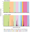
Extended data Figure 2. Glycan microarray analyses of murine Fc-MelLec and human Langerin.
These 496 lipid-linked probes are arranged according to their backbone sequences as annotated in the coloured panels below the figure. Lac, lactose; LacNAc, N-acetyllactosamine; LNnT, lacto-N-neotetraose; LNT, lacto-N-tetraose; PolyLac, polylactosamine; GAGs, glycosaminoglycans; Misc., miscellaneous. The signals are means of fluorescence intensities of duplicate spots, printed at 5 fmol per spot level with error bars representing half of the difference between the two values. The signals shown together with the probe sequences are in Supplementary Table 1. ns, not significant.
Extended data Figure 3
Extended data Figure 3. MelLec recognises DHN-melanin.
a, Representative immunofluorescence micrographs using Fc-MelLec as a probe showing the surface distribution of MelLec-ligands on A. fumigatus wild-type and a variety of melanin deficient mutants, as indicated. Lower panels show conidia treated with 1M NaOH. b, Representative immunofluorescence and light micrograph images of ΔrodAΔpksP A. fumigatus conidia stained with Fc-MelLec or ConA-FITC, as indicated. c, Representative histograms showing the presence or absence of MelLec or Dectin-1 ligands on ΔrodA or ΔrodAΔpksP A. fumigatus conidia, as indicated. Fungal particles were stained with Fc-MelLec (red) or Fc-Dectin-1 (green) and analysed by flow cytometry. Grey histograms indicate secondary only control. d, Representative histogram showing the presence Dectin-1 ligands on ΔpksP A. fumigatus conidia. Fungal particles were stained with Fc-Dectin-1 (green) or Fc-CLEC12b (Fc-control; blue) and analysed by flow cytometry. e, Representative histogram showing the presence MelLec ligands on melanin ghosts of A. fumigatus conidia. Fungal particles were stained with Fc-MelLec (red) or Fc-CLEC12b (Fc-control; blue) and analysed by flow cytometry. a-e, Experiments were repeated 3 times independently, with similar results. f, Flow cytometric analysis of melanised (red) and non-melanised (grey) Cryptococcus neoformans yeast and melanin ghosts, and B16 melanoma cells, stained with Fc-MelLec, as indicated. Experiment was repeated independently twice, with similar results.
Extended data Figure 4
Extended data Figure 4. MelLec recognises naphthalene-diol.
a, Detection of MelLec ligands in ghosts of melanin mutants of A. fumigatus by ELISA, as indicated. Values show mean ± SD. b, Representative immunofluorescence and light micrograph images of Fc-MelLec ligands on NaOH-treated A. fumigatus B5233 conidia following pre-treatment with ghosts of ΔpksP or Δayg1 conidia, as indicated. c, Representative immunofluorescence and light micrograph images of MelLec ligands on NaOH-treated wild-type A. fumigatus conidia following pre-treatment with or without heptaketide naphthopyrone (YWA1). a-c, Experiments were repeated at least 3 times independently, with similar results. d, Detection of Fc-MelLec or Fc-Dectin-1 ligands on ΔrodA conidia following pre-treatment with (blue) or without (red) heptaketide naphthopyrone (YWA1). Experiment was repeated independently twice, with similar results. Detection of 1,8-DHN (e) and 1,2-DHN and 1,4-DHN (f) by Fc-MelLec and Fc-control using ELISA, as indicated. Values show mean ± SD. g, Detection of 1,8-DHN, naphthalene and 1-naphthol by Fc-MelLec using ELISA, as indicated. Values show mean ± SD. e-g, Experiments were repeated at least 3 times independently, with similar results.
Extended data Figure 5
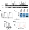
Extended data Figure 5. MelLec is expressed at the cell surface.
a, RT-PCR detection of MelLec expression in various tissues, as indicated. The expression of glycerol-3-phosphate dehydrogenase (G3PDH) in these samples, also used for the characterization of MCL, is shown as a control. Experiment was performed once (for gel source data, see Supplementary Figure 1). b, Flow cytometric analysis of surface expression of HA-tagged murine and human MelLec on the surface of NIH3T3 fibroblasts (black open histograms). NIH3T3 cells transfected with vector only served as controls (grey filled histograms). c, Western blot analysis of lysates of HA-tagged mMelLec expressing NIH3T3 cells under reducing and non-reducing conditions and with and without N-glycosidase. HA-tagged mCLEC12A expressing NIH3T3 cells served as controls (for blot source data, see Supplementary Figure 1). d, Relative binding of FITC-labelled ΔrodA A. fumigatus conidia to NIH3T3 cells transduced with vector only, Dectin-1 or MelLec, as determined by flow cytometry. Values shown are mean ± SD, analysed by one-way ANOVA. e, Screening of hybridoma supernatants on mMelLec-expressing (red) and parental (black) NIH3T3 cells. b-e, Experiments were repeated at least 3 times independently, with similar results. *, p≤0.05; Ns, not significant.
Extended data Figure 6
Extended data Figure 6. Murine MelLec is not expressed by myeloid cells.
Flow cytometric analysis of MelLec expression on various ex vivo and in vitro derived myeloid cells (a) and peripheral blood, bone marrow, lymph nodes and spleen (b), as indicated. a-b, Experiments were repeated at least twice independently, with similar results. c, Flow cytometric analysis of MelLec expression on CD61+ platelets. Experiment was repeated at least 3 times independently, with similar results. BM, bone marrow; DC, dendritic cell; mø, macrophage; LPS, lipopolysaccharide.
Extended data Figure 7
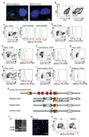
Extended data Figure 7. MelLec expression in tissues and generation of MelLec-/- mice.
a, Immunofluorescence microscopy of MelLec-expressing versus control NIH3T3 cells labelled with αMelLec (green). Nuclei are stained with DAPI (blue). Experiments were repeated at least 3 times independently, with similar results. b, Exemplar flow cytometric gating strategy for identification of live cells from tissue. c, Flow cytometric analysis of MelLec expression on live CD45- CD31+ EpCAM- and EpCAM+ populations in the liver, as indicated. Flow cytometric analysis of MelLec expression on live CD45- CD31+ cells in the heart (d), kidney (e) and small intestine (f). Flow cytometric analysis of MelLec expression on live CD45-EpCAM+ cells in the heart (g), kidney (h), small intestine (i) and epidermis (j). b-j Experiments were repeated at least twice independently, with similar results. Black lines, isotype controls. k, Schematic representation of the wild-type CLEC1A locus, gene targeting vector, PCR primer sites, and correctly targeted recombinant gene-targeted allele. l, PCR analysis of gene-targeted mice (for gel source data, see Supplementary Figure 1). +/+, wild-type, +/- heterozygous and -/- homozygous for the targeted allele. m, Immunofluorescence microscopy of naïve lung tissue from MelLec-/- mice (labelling of wild-type lung is shown in Fig. 3b). n, Analysis of MelLec expression in disaggregated lung tissue from wild-type (wt) or MelLec-/- mice by flow cytometry. l-n, Experiments were repeated at least 3 times independently, with similar results.
Extended data Figure 8
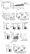
Extended data Figure 8. MelLec-/- mice show early inflammatory defects upon challenge with A. fumigatus.
a, Survival (left) and weight measurements (right) of immunocompetent mice following intratracheal (i.t.) infection with 107 A. fumigatus conidia (n=4 animals per group). Values shown are mean ± SD. b, Exemplar flow cytometric gating strategy for identification of CD45+ cells from BAL. c, Representative FACS profiles of pulmonary CD11b+ Ly6Ghigh neutrophils in wild-type and MelLec-/- mice 4 h after challenge with 107 A. fumigatus conidia (wt n=29 animals; MelLec-/- n=26 animals). Pulmonary CD11b+ Ly6Ghigh neutrophils (wt n=29 animals, MelLec-/- n=26 animals) (d) and cytokines (n=25 animals per group) (e) in mice 4 h after challenge with 107 A. fumigatus conidia, as indicated. f, Pulmonary CD11b+ Ly6Ghigh neutrophils in mice 4 h after challenge with melanin ghosts (160 µg) of A. fumigatus (wt n=15 animals; MelLec-/- n=10 animals). Samples with blood contamination were excluded. g, Pulmonary CD11b+ Ly6Ghigh neutrophils in mice 24 h after challenge with 107 A. fumigatus conidia (wt n=33 animals, MelLec-/- n=30 animals). h, Cellular inflammatory profiles of mice after 24 h challenge with 107 A. fumigatus conidia, as indicated (wt n=33 animals, MelLec-/- n=30 animals). Alveolar macrophages were defined as CD11c+ Siglec-F+, inflammatory macrophages as CD11b+ F4/80+ and eosinophils as CD11b+ Siglec-F+. d-h, Values shown are mean ± SEM of pooled data from at least two independent experiments, analysed by two-sided Mann-Whitney U test. i, Expression of MelLec on pulmonary CD45- CD31+ cells isolated from uninfected mice (black) and mice 24 h after infection with A. fumigatus conidia (red). Grey line, isotype control (n=3 animals per group). j, Pulmonary CD11b+ Ly6Ghigh neutrophils in mice 4 h after challenge with 107 ΔpksP A. fumigatus conidia (wt n=7 animals, MelLec-/- n=8 animals). Values shown are mean ± SEM of pooled data from two independent experiments, analysed by two-sided Mann-Whitney U test. k, Representative FACS profiles of pulmonary CD11b+ Ly6Ghigh neutrophils in wild-type and MelLec-/- mice 4 h after challenge with 107 ΔpksP A. fumigatus conidia (wt n=7 animals, MelLec-/- n=8 animals). *, p≤0.05; ns, not significant.
Extended data Figure 9
Extended data Figure 9. MelLec-/- mice show alterations in anti-fungal immunity during systemic infection.
a, Survival of corticosteroid treated mice following intratracheal (i.t.) infection with 105 A. fumigatus conidia (n=15 animals per group). Pooled data from two independent experiments, analysed by log-rank test. b, Fungal burdens in various mouse tissues, as indicated, 4 days after i.v. infection with 106 A. fumigatus conidia (n=12 animals per group). Values shown are mean ± SEM of pooled data from two independent experiments, analysed by two-sided Mann-Whitney U test. c, Tissue section of kidney from day 4 infected MelLec-/- mouse stained with Grocott's methenamine silver stain and haematoxylin (n=3 animals per group). d, Immunofluorescence microscopy of brain from day 2 infected MelLec-/- mouse (n=3 animals per group). Fungi are stained with calcofluor white (blue), leukocytes with Gr1 (green), and DNA with propidium iodide (red). e, Fungal burdens in various mouse tissues, as indicated, 4 days after i.v. infection with 106 ΔpksP A. fumigatus conidia (n=4 animals per group). Values shown are mean ± SD, analysed by two-sided Mann-Whitney U test. *, p≤0.05; ns, not significant.
Extended data Figure 10
Extended data Figure 10. A SNP in human MelLec influences anti-Aspergillus inflammatory responses.
a, Representative histograms showing the presence or absence of human MelLec ligands on ΔrodA or ΔrodAΔpksP A. fumigatus conidia, as determined by flow cytometry. Fc-CLEC12b was used as a control (Fc-control). Experiment was repeated at least 3 times independently, with similar results. b, Survival of irradiated wild-type mice reconstituted with wild-type or MelLec-/- bone-marrow (BM), as indicated, following i.v. infection with 106 A. fumigatus conidia (n=16 animals per group). Pooled data from two independent experiments, analysed by log-rank test. c, Inflammatory cytokine production in monocyte-derived macrophages isolated from genotyped individuals, following stimulation with LPS (wild-type n=14 individuals, SNP allele n=5 individuals). Values shown are mean ± SD, analysed by two-sided Mann-Whitney U test. d, Inflammatory cytokine production in PBMCs isolated from genotyped Dutch individuals, following stimulation with heat-killed A. fumigatus conidia (wild-type n=72 individuals, SNP allele n=17 individuals). Boxes represent the median values and interquartile ranges; whiskers represent minimum and maximum values, analysed by two-sided Mann-Whitney U test. e, Inflammatory cytokine production in transduced RAW264.7 macrophages expressing wild-type or SNP allele following stimulation with ΔrodA or ΔrodAΔpksP A. fumigatus conidia, as indicated. Values shown are mean ± SD, analysed by one-way ANOVA and repeated at least 3 times independently, with similar results. *, p≤0.05; ns, not significant.
Figure 1
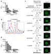
Figure 1. MelLec recognises selected fungi.
Representative histograms showing (a) A. fumigatus conidia and germlings (cultured for 8 h at 37°C), or (b) yeasts of C. albicans and S. cerevisiae, and conidia of F. pedrosoi, stained with Fc-MelLec or Fc-CLEC12b (Fc-control) and analysed by flow cytometry. Representative light microscope images and immunofluorescence micrographs using Fc-MelLec to detect ligands on A. fumigatus following conidial swelling and germination over time, as indicated (c), following treatment with 1M NaOH (d), and on rodlet-deficient (ΔrodA) conidia (e). a-e, Experiments were repeated at least 3 times independently, with similar results.
Figure 2
Figure 2. MelLec recognises DHN-melanin.
a, Detection of MelLec ligands in alkali insoluble or soluble A. fumigatus cell wall fractions by ELISA. Values show mean ± SD. b, Representative histograms showing ΔrodA or ΔpksP A. fumigatus conidia, stained with Fc-MelLec or Fc-CLEC12b (Fc-control) and analysed by flow cytometry. c, The biosynthetic pathway of DHN-melanin (left) and representative immunofluorescence micrographs using Fc-MelLec to detect ligands on A. fumigatus strains deficient in these enzymes (right). The conidial rodlet layer was removed with 1M NaOH prior to staining. d, Detection of YWA1 by Fc-MelLec and Fc-control by ELISA. Values show mean ± SD. a-d, Experiments were repeated at least 3 times independently, with similar results.
Figure 3
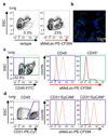
Figure 3. MelLec is expressed on non-myeloid cells in mouse.
a, Analysis of disaggregated lung tissue by flow cytometry with αMelLec. b, Immunofluorescence microscopy of lung tissue stained with αMelLec (green). Nuclei are stained with DAPI (blue). Flow cytometric analysis of MelLec expression on live CD45+ and CD45- cells (c), and CD45- CD31+ EpCAM- and EpCAM+ cells (d) in the lung. a-d, Experiments were repeated at least 3 times independently, with similar results.
Figure 4
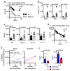
Figure 4. MelLec is required to prevent disseminated infection in mice and humans.
a, Survival of mice following intravenous (i.v.) infection with 106 A. fumigatus conidia (n=16 animals per group). Pooled data from two independent experiments, analysed by log-rank test. Tissue fungal burdens (b) and brain cytokine levels (c) of mice 4 days after i.v. infection with 106 A. fumigatus conidia (n=12 animals per group). b-c, Values shown are mean ± SEM of pooled data from two independent experiments, analysed by two-sided Mann-Whitney U test. d, Survival of mice following i.v. infection with 106 ΔpksP A. fumigatus conidia (n=15 animals per group). Pooled data from two independent experiments, analysed by log-rank test. e, Cumulative incidence analysis of invasive aspergillosis after transplantation according to donor (wildtype, n=238 individuals; SNP allele, n=72 individuals) or recipient (wildtype, n=228 individuals; SNP allele, n=80 individuals) CLEC1A rs2306894 genotypes, analysed by two-sided Gray’s test. f, Cytokine production in monocyte-derived macrophages, following stimulation with A. fumigatus conidia (wildtype, n=14 individuals; SNP allele, n=5 individuals). Values shown are mean ± SD, analysed by two-sided Mann-Whitney U test. *, p≤0.05; ns, not significant; HR, hazard ratio.

Comment in

References

    1. Hardison SE, Brown GD. C-type lectin receptors orchestrate antifungal immunity. Nat Immunol. 2012;13:817–822. - PMC - PubMed
    1. Nosanchuk JD, Casadevall A. The contribution of melanin to microbial pathogenesis. Cell Microbiol. 2003;5:203–223. - PubMed
    1. Heinekamp T, et al. Aspergillus fumigatus melanins: interference with the host endocytosis pathway and impact on virulence. Front Microbiol. 2012;3:440. - PMC - PubMed
    1. Pyz E, Brown GD. Screening for ligands of C-type lectin-like receptors. Methods Mol Biol. 2011;748:1–19. - PubMed
    1. Colonna M, Samaridis J, Angman L. Molecular characterization of two novel C-type lectin-like receptors, one of which is selectively expressed in human dendritic cells. Eur J Immunol. 2000;30:697–704. - PubMed

Publication types

MeSH terms