

Development of tissue inflammation accompanied by NLRP3 inflammasome activation in rabbits infected with Treponema pallidum strain Nichols
- PMID: 29490620
- PMCID: PMC5831842
- DOI: 10.1186/s12879-018-2993-0
Development of tissue inflammation accompanied by NLRP3 inflammasome activation in rabbits infected with Treponema pallidum strain Nichols
Abstract
Background: The inflammasome responses in Treponema pallidum infection have been poorly understood to date. This study aimed to investigate the expression of the nucleotide-binding leucine-rich receptor protein 3 (NLRP3) inflammasome in the development of tissue inflammation in rabbits infected with T. pallidum.
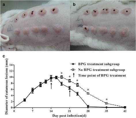
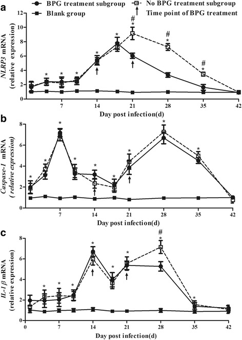
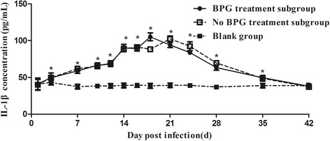
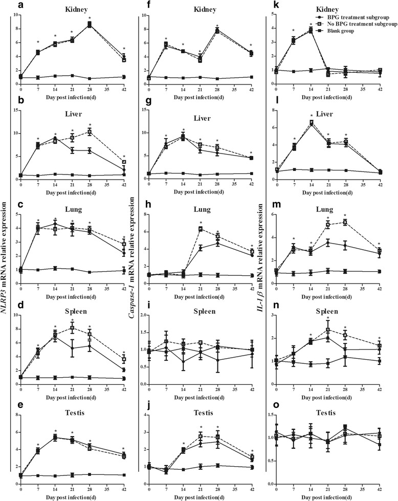
Methods: Forty-five rabbits were randomly assigned to a blank group or an infection group, and the latter was divided into no benzathine penicillin G (BPG) and BPG treatment subgroups. Rabbits in the infection group were injected intradermally with 0.1 mL of a 107/mL T. pallidum suspension at 10 marked sites along the back, and the blank group was treated with normal saline. The BPG treatment subgroup received 200,000 U of BPG administered intramuscularly twice, at 14 d and 21 d post-infection. The development of lesions was observed, and biopsies of the injection site and various organs, including the kidney, liver, spleen, lung, and testis, were obtained for NLRP3, caspase-1, and interleukin-1β (IL-1β) mRNA analysis during infection. Blood was also collected for the determination of IL-1β concentration.
Results: Rabbits infected with T. pallidum (both the BPG treatment and no BPG treatment subgroups), exhibited NLRP3 inflammasome activation and IL-1β secretion in cutaneous lesions, showing a trend in elevation to decline; NLRP3 mRNA expression reached a peak at 18 d in the BPG treatment subgroup and 21 d in the no BPG treatment subgroup and returned to "normal" levels [vs. the blank group (P > 0.05)] at 42 d post-infection. The trend was similar to the change in cutaneous lesions in the infected rabbits, which reached a peak at 16 d in the BPG treatment subgroup and 18 d in the no BPG treatment subgroup. NLRP3, caspase-1, and IL-1β mRNA expression levels were slightly different in different organs. NLRP3 inflammasome activation was also observed in the kidney, liver, lung, spleen and testis. IL-1β expression was observed in the kidney, liver, lung and spleen; however, there was no detectable level of IL-1β in the testes of the infected rabbits.
Conclusions: This study established a clear link between NLRP3 inflammasome activation and the development of tissue inflammation in rabbits infected with T. pallidum. BPG therapy imperceptibly adjusted syphilitic inflammation.
Keywords: Il-1β; Inflammation; NLRP3; Rabbit; Treponema pallidum.
Conflict of interest statement
Ethics approval
This study was approved by the animal experimental ethics committee of the Medical College of Xiamen University.
Consent for publication
Not applicable.
Competing interests
The authors declare that they have no competing interests.
Publisher’s Note
Springer Nature remains neutral with regard to jurisdictional claims in published maps and institutional affiliations.
Figures
References
Publication types
MeSH terms
Substances
Grants and funding
- 81772260/the National Natural Science Foundation/International
- 81672094/the National Natural Science Foundation/International
- 81471231/the National Natural Science Foundation/International
- 81101324/the National Natural Science Foundation/International
- 81171625/the National Natural Science Foundation/International
- 81771312/the National Natural Science Foundation/International
- 81471967/the National Natural Science Foundation/International
- 81401749/the National Natural Science Foundation/International
- 81301501/the National Natural Science Foundation/International
- 81201360/the National Natural Science Foundation/International
- 81271335/the National Natural Science Foundation/International
- 2013-ZQN-ZD-35/the Key Project of Cultivating Young Talent in Fujian Province's Health System/International
- 2014-ZQN-ZD-34/the Key Project of Cultivating Young Talent in Fujian Province's Health System/International
- 2014D001/the National Science Foundation for Distinguished Young Scholars of Fujian/International
- 2017-2-105/the Youth Foundation Project of Fujian Provincial Health Department/International
- 2017-2-113/the Youth Foundation Project of Fujian Provincial Health Department/International
- 2014-2-68/the Youth Foundation Project of Fujian Provincial Health Department/International
- 2014-CXB-40/the Medical Innovation Project of Fujian Health Development Planning Commission/International
- 2012-CXB-33/the Medical Innovation Project of Fujian Health Development Planning Commission/International
- 3502Z20159016/the major special projects of serious illness in Xiamen/International
- 2016J01628/the Natural Science Foundation of Fujian Province/International
- 2018D0014/the Natural Science Foundation of Fujian Province/International
LinkOut - more resources
Full Text Sources
Other Literature Sources
Medical
