. 2018 Mar 1;18(1):9.
doi: 10.1186/s40644-018-0143-y.

Imaging features of fibrolamellar hepatocellular carcinoma in gadoxetic acid-enhanced MRI

Affiliations

Imaging features of fibrolamellar hepatocellular carcinoma in gadoxetic acid-enhanced MRI

Viktoria Palm et al. Cancer Imaging. .

Abstract

Background: Fibrolamellar hepatocellular carcinoma (FLC) is a rare malignancy occurring in young patients without cirrhosis. Objectives of our study were to analyze contrast material uptake in hepatobiliary phase imaging (HBP) in gadoxetic acid-enhanced liver MRI in patients with FLC and to characterize imaging features in sequence techniques other than HBP.

Methods: In this retrospective study on histology-proven FLC, contrast material uptake in HBP was quantitatively assessed by calculating the corrected FLC enhancement index (CEI) using mean signal intensities of FLC and lumbar muscle on pre-contrast imaging and HBP, respectively. Moreover, enhancement patterns in dynamic contrast-enhanced MRI and relative signal intensities compared with background liver parenchyma were determined by two radiologists in consensus for HBP, diffusion-weighted imaging using high b-values (DWI), and T2 and T1 weighted pre-contrast imaging.

Results: In 6 of 13 patients with FLC gadoxetic acid-enhanced liver MRI was available. The CEI suggested presence of HBP contrast material uptake in all FLCs. A mean CEI of 1.35 indicated FLC signal increase of 35% in HBP compared with pre-contrast imaging. All FLCs were hypointense in HBP compared with background liver parenchyma. Three of 6 FLCs had arterial hyperenhancement and venous wash-out. In DWI and T2 weighted imaging, 5 of 6 FLCs were hyperintense. In T1 weighted imaging, 5 of 6 FLCs were hypointense.

Conclusion: Hepatobiliary uptake of gadoxetic acid was quantitatively measurable in all FLCs investigated in our study. The observation of hypointensity of FLCs in HBP compared with background liver parenchyma emphasizes the role of gadoxetic acid-enhanced liver MRI for non-invasive diagnosis of FLC and its importance in the diagnostic work-up of indeterminate liver lesions.

Keywords: Contrast media; Delayed diagnosis; Diagnostic imaging; Liver neoplasms; Magnetic resonance imaging.


Conflict of interest statement

Ethics approval and consent to participate

Ethical approval was obtained by the local institutional review board (Ethical Committee of the University of Heidelberg, S-043/2011).

Consent for publication

Not applicable

Competing interests

The authors declare that they have no competing interests.

Publisher’s Note

Springer Nature remains neutral with regard to jurisdictional claims in published maps and institutional affiliations.

Figures
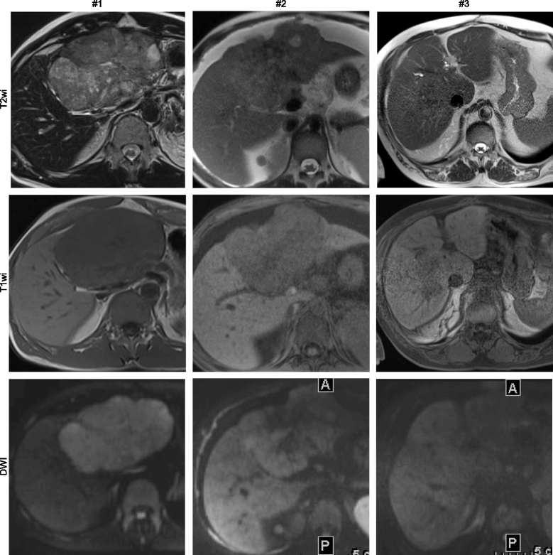
Fig. 1
Fig. 1
Image panel of patients #1, #2, and #3 displaying representative sections through individual fibrolamellar carcinoma in non-contrast enhanced techniques including T2 weighted imaging (T2wi), T1 weighted imaging (T1wi), and diffusion weighted imaging using high b-values (DWI)
Fig. 2
Fig. 2
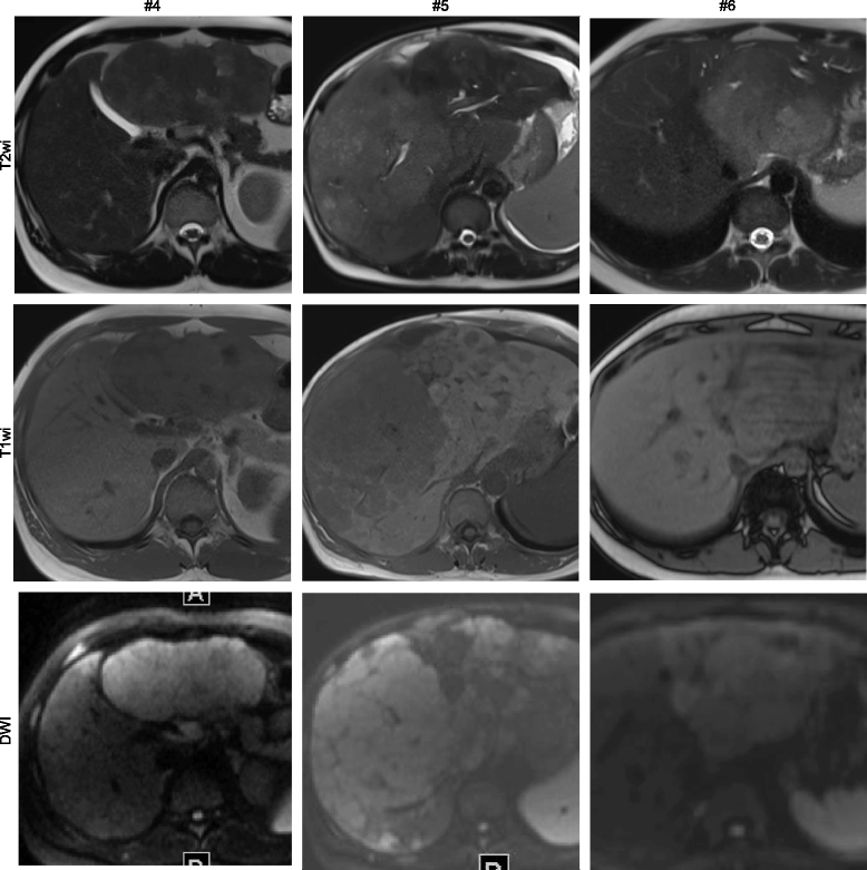
Image panel of patients #4, #5, and #6 displaying representative sections through individual fibrolamellar carcinoma in non-contrast enhanced imaging techniques including T2 weighted imaging (T2wi), T1 weighted imaging (T1wi), and diffusion weighted imaging using high b-values (DWI)
Fig. 3
Fig. 3
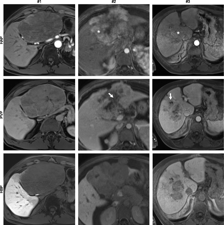
Image panel of patients #1, #2, and #3 displaying representative sections through individual fibrolamellar carcinoma in contrast enhanced imaging techniques including hepatic arterial phase T1 weighted imaging (HAP), portal venous phase T1 weighted imaging (PVP), and hepatobiliary phase T1 weighted imaging (HBP). In patients with history of TACE prior to gadoxetic acid enhanced MRI (#2, #3) areas of non-viable tumor are indicated in the portal venous phase (arrows) and areas of viable tumor are indicated in the hepatic arterial phase (star)
Fig. 4
Fig. 4
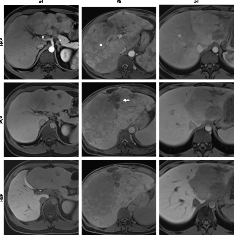
Image panel of patients #1, #2, and #3 displaying representative sections through individual fibrolamellar carcinoma in contrast enhanced imaging techniques including hepatic arterial phase T1 weighted imaging (HAP), portal venous phase T1 weighted imaging (PVP), and hepatobiliary phase T1 weighted imaging (HBP). In patients with history of TACE prior to gadoxetic acid enhanced MRI (#5) areas of non-viable tumor are indicated in the portal venous phase (arrows) and areas of viable tumor are indicated in the hepatic arterial phase (star)
Fig. 5
Fig. 5
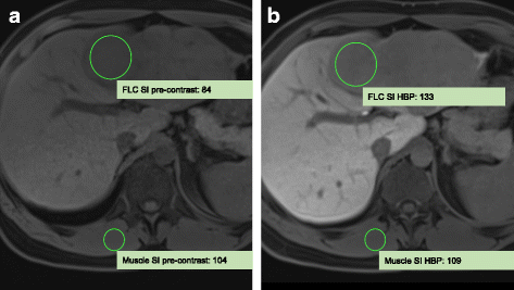
Assessment of the corrected enhancement index (CEI) in patient #4. a shows the pre-contrast scan. b shows the hepatobiliary phase. CEI is 1.51 indicating a signal increase of 51% normalized to lumbar muscle signal intensity. FLC, fibrolamellar carcinoma; HBP, hepatobiliary phase; SI, signal intensity

References

    1. Torbenson M. Fibrolamellar carcinoma: 2012 update. Scientifica (Cairo) Hindawi. 2012;2012:743790–743715. - PMC - PubMed
    1. Craig JR, Peters RL, Edmondson HA, Omata M. Fibrolamellar carcinoma of the liver: a tumor of adolescents and young adults with distinctive clinico-pathologic features. Cancer. 1980;46:372–379. doi: 10.1002/1097-0142(19800715)46:2<372::AID-CNCR2820460227>3.0.CO;2-S. - DOI - PubMed
    1. Honeyman JN, Simon EP, Robine N, Chiaroni-Clarke R, Darcy DG, Lim IIP, et al. Detection of a recurrent DNAJB1-PRKACA chimeric transcript in fibrolamellar hepatocellular carcinoma. Science. 2014;343:1010–1014. doi: 10.1126/science.1249484. - DOI - PMC - PubMed
    1. Graham RP, Jin L, Knutson DL, Kloft-Nelson SM, Greipp PT, Waldburger N, et al. DNAJB1-PRKACA is specific for fibrolamellar carcinoma. Mod Pathol. 2015;28:822–829. doi: 10.1038/modpathol.2015.4. - DOI - PubMed
    1. Corrigan K, Semelka RC. Dynamic contrast-enhanced MR imaging of fibrolamellar hepatocellular carcinoma. Abdom Imaging. 1995;20:122–125. doi: 10.1007/BF00201518. - DOI - PubMed

Supplementary concepts