. 2018 Mar 1;13(1):11.
doi: 10.1186/s13024-018-0244-x.

Early long-term administration of the CSF1R inhibitor PLX3397 ablates microglia and reduces accumulation of intraneuronal amyloid, neuritic plaque deposition and pre-fibrillar oligomers in 5XFAD mouse model of Alzheimer's disease

Affiliations

Early long-term administration of the CSF1R inhibitor PLX3397 ablates microglia and reduces accumulation of intraneuronal amyloid, neuritic plaque deposition and pre-fibrillar oligomers in 5XFAD mouse model of Alzheimer's disease

Justyna Sosna et al. Mol Neurodegener. .

Abstract

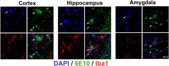
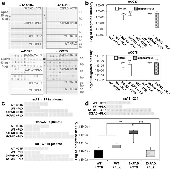
Background: Besides the two main classical features of amyloid beta aggregation and tau-containing neurofibrillary tangle deposition, neuroinflammation plays an important yet unclear role in the pathophysiology of Alzheimer's disease (AD). Microglia are believed to be key mediators of neuroinflammation during AD and responsible for the regulation of brain homeostasis by balancing neurotoxicity and neuroprotective events. We have previously reported evidence that neuritic plaques are derived from dead neurons that have accumulated intraneuronal amyloid and further recruit Iba1-positive cells, which play a role in either neuronal demise or neuritic plaque maturation or both.

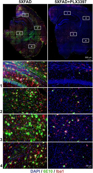
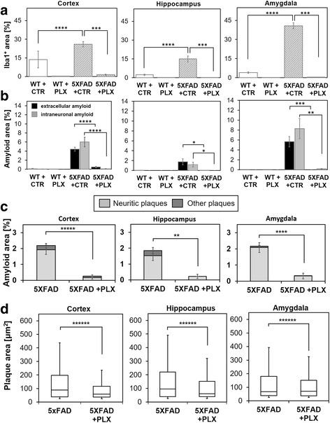
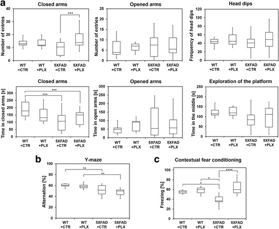
Methods: To study the impact of microglia on neuritic plaque development, we treated two-month-old 5XFAD mice with a selective colony stimulation factor 1 receptor (CSF1R) inhibitor, PLX3397, for a period of 3 months, resulting in a significant ablation of microglia. Directly after this treatment, we analyzed the amount of intraneuronal amyloid and neuritic plaques and performed behavioral studies including Y-maze, fear conditioning and elevated plus maze.

Results: We found that early long-term PLX3397 administration results in a dramatic reduction of both intraneuronal amyloid as well as neuritic plaque deposition. PLX3397 treated young 5XFAD mice also displayed a significant decrease of soluble fibrillar amyloid oligomers in brain lysates, a depletion of soluble pre-fibrillar oligomers in plasma and an improvement in cognitive function measured by fear conditioning tests.

Conclusions: Our findings demonstrate that CSF1R signaling, either directly on neurons or mediated by microglia, is crucial for the accumulation of intraneuronal amyloid and formation of neuritic plaques, suggesting that these two events are serially linked in a causal pathway leading to neurodegeneration and neuritic plaque formation. CSF1R inhibitors represent potential preventative or therapeutic approach that target the very earliest stages of the formation of intraneuronal amyloid and neuritic plaques.

Keywords: Alzheimer’s disease; Amyloid beta; Intraneuronal amyloid; Microglia; Neuroinflammation; Plaques.


Conflict of interest statement

Ethics approval and consent to participate

All mouse experiments were performed according to animal protocols approved by the Institutional Animal Care and Use Committee at the University of California, Irvine.

Consent for publication

The authors have no conflict of interest.

Competing interests

The authors declare that they have no competing interests.

Publisher’s Note

Springer Nature remains neutral with regard to jurisdictional claims in published maps and institutional affiliations.

Figures

Fig. 1
Fig. 1
Intraneuronal amyloid and neuritic plaques are present in 5 mo 5XFAD mice. Representative 63× images of intraneuronal amyloid (asterisk), neuritic plaques (arrows) with DAPI-positive core (blue) surrounded by a 6E10-positive amyloid rim (green) and Iba1-positive cells (red) in cortex, hippocampus and amygdala. Arrowheads point to non-neuritic 6E10-positive plaques without a DAPI-positive center. The confocal acquisition was applied and a total z-plane of 10 μm was merged into single overlay images. Bar = 30 μm
Fig. 2
Fig. 2
Long-term administration of PLX3397 ablates microglia and reduces neuritic plaque formation in 5XFAD mice. Top panels: representative 20× images of coronal brain sections with immunolabeling of microglia (Iba1, red) and staining for amyloid Aβ (6E10, green) in five-month-old 5XFAD mice kept on a standard diet or fed for 3 months with PLX3397 (290 mg/kg). Bottom panels: representative images of magnified regions of (1) hippocampus, (2) cortex, (3) thalamus and (4) amygdalar nucleus. The confocal acquisition was applied and a total z-plane of 30 μm was scanned across all regions and merged into single overlay images
Fig. 3
Fig. 3
PLX3397 treatment reduces microglia, accumulation of intraneuronal amyloid and extracellular amyloid deposits in 5XFAD mice. a Area occupied by Iba1-positive microglia in the analyzed brains is increased by ~ 40%–90% in the regions of cortex, hippocampus and amygdala in 5XFAD mice compared to wild-type (p < 0.0001 denoted by ****). PLX3397 treatment eliminates ~ 99% of microglia in both wild-type and 5XFAD mice (p < 0.001 denoted by ***). Graph bars indicate mean ± SEM (n = 9/group). b Area fraction occupied by intraneuronal amyloid and extracellular amyloid deposits was quantified for the regions of cortex, hippocampus and amygdala independently. Long-term treatment with PLX3397 significantly prevented accumulation of the intraneuronal amyloid and nearly completely aborted formation of amyloid plaques in the cortex (p < 0.0001 denoted by ****). In the hippocampus, the accumulation of intraneuronal amyloid was significantly reduced (p < 0.024 denoted by *) as the formation of extracellular amyloid plaques (p < 0.05 denoted by *). In the amygdala region of the brains intraneuronal amyloid was reduced (p < 0.0002 denoted by ***) and formation of amyloid plaques was prevented (p < 0.003 denoted by **). Graph bars indicate mean ± SEM (n = 9/group). c Based on morphology and presence or absence of DAPI-positive core, extracellular amyloid was divided into neuritic or other types of plaques and quantified in brains. Treatment with PLX3397 significantly reduced area fraction occupied by neuritic plaques and other types of plaques (non-neuritic) in the cortex (p < 0.000005 denoted by *****), hippocampus (p < 0.005 denoted by **) and amygdala (p < 0.00001 denoted by ****). Graph bars indicate mean ± SEM (n = 12/group). d Size of amyloid plaques was quantified in three regions of brains. Treatment with PLX3397 significantly reduced the size of amyloid plaques in the cortex, hippocampus and amygdala (p < 0.000001, denoted by ******). Graph bars indicate mean ± SEM (n = 12/group and the total size of > 300 plaques/group were analyzed)
Fig. 4
Fig. 4
PLX3397 treatment improves behavioral phenotype. Contextual fear conditioning (a), Y-maze (b) and elevated plus maze (c) tests were performed. a In the contextual fear conditioning test, 5XFAD mice show significantly lower levels of contextual freezing than wild-type controls (p < 0.02, denoted by *). However, treatment with PLX3397 significantly reversed the impaired fear behavior to the level of a normal reconsolidation of the contextual fear memory (p < 0.00008 with Tukey correction test, denoted by ****). b The total number of arm entries was significantly changed in the group of 5XFAD mice regardless the treatment (p < 0.005 with Tukey correction test, denoted by **), indicating lower levels of motor and exploratory activity in the group of AD mice. c In the elevated plus maze test, although the number of entries into closed arms was increased in the 5XFAD mice treated with PLX3397 (p < 0.001 denoted by ***), the number of entries into opened arms was not significantly different among all four groups. Neither another behavioral parameter such as head dips, which is a complementary parameter to assess risky and or an exploratory behavior in the evaluation of anxiety, was not changed between groups. Whereas, 5XFAD mice spend less time in the closed arms (p < 0.0002, denoted by ***), regardless the treatment, there was no difference in the time spent in the opened arms or exploring the center zone of the platform. Box plots show median with interquartile lower and upper range and the minimum and maximum values. Total number of wild-type animals in the behavioral study was four animals (two males and two females) with drug and six animals without drug (three males and three females), respectively. The total number of animals in the behavioral study was 12 5XFAD animals (five males and seven females) with drug and 11 5XFAD animals without drug (six males and five females), respectively
Fig. 5
Fig. 5
Treatment with PLX3397 reduces the level of fibrillar amyloid in the total brain lysates and pre-fibrillar oligomers in the blood plasma of 5XFAD mice. a An equal amount (6 μg) of total lysate from the cortex (cx) or hippocampus (hp) of each group of mice (male and female) were spotted on the membrane and incubated with antibodies specifically detecting pre-fibrillar oligomers (mA11) or fibrillar aggregates (mOC). 5 μM of oligomerized for 3 days Aβ42 was spotted as a control for antibodies to confirm the ability of the antibodies to distinguish between oligomeric vs. fibrillar conformation. Additionally, 5/10 μg of rabbit IgG were spotted as a positive control for the secondary anti-rabbit antibody. b The dot blot analyses were quantified for the mOC23 and mOC78. Whereas wild-type brains were free of amyloid, the fibrillar soluble amyloid was increased in the cortex and hippocampal lysates. However, long-term treatment with PLX3397 (290 mg/kg) decreased significantly the amount of soluble fibrillar mOC23 amyloid in the cortex (p < 0.004 with Tukey test, denoted by **) and in the hippocampus (p < 0.05 with Tukey test, denoted by *) and soluble fibrillar mOC78 amyloid in the cortex (p < 0.0018 with Tukey test, denoted by **) and in the hippocampus (p < 0.0012, denoted by **) when compared with the control-chow treated 5XFAD mice. c An equal amount of blood plasma (1 μg) of each group (male and female) was spotted on the membrane and incubated as in (a). Neither the antibodies against fibrillar mOC23, mOC78 nor mA11–118 against pre-prefibrillar oligomers detected a soluble amyloid in blood plasma. However, antibody mA11–204 has detected pre-fibrillar amyloid in the blood plasma of analyzed cohorts. d Quantification of the dot blot for mA11–204 presence in blood plasma. Some endogenous pre-fibrillar amyloid was detected in the wild-type animals, however, the amount of mA11–204-positive amyloid increased significantly in the plasma of 5XFAD animals (p < 0.0011 with Tukey test, denoted by **). Long-term administration of PLX3397 in young 5XFAD decreased the amount of pre-fibrillar oligomers (mA11–204) in blood plasma (p < 0.00082 with Tukey test, denoted by ***). A-D each assay was performed once for each individual animal due to non-significant inter-assay variability

References

    1. Ramirez-Bermudez J. Alzheimer's disease: critical notes on the history of a medical concept. Arch Med Res. 2012;43(8):595–599. doi: 10.1016/j.arcmed.2012.11.008. - DOI - PubMed
    1. Alzheimer A. Über eine eigenartige Erkrankung der Hirnrinde. Allg Z Psychiatrie Psychisch-gerichtl Med. 1907;64:146–148.
    1. Barage SH, Sonawane KD. Amyloid cascade hypothesis: pathogenesis and therapeutic strategies in Alzheimer's disease. Neuropeptides. 2015;52:1–18. doi: 10.1016/j.npep.2015.06.008. - DOI - PubMed
    1. Doody RS, Raman R, Farlow M, Iwatsubo T, Vellas B, Joffe S, Kieburtz K, He F, Sun X, Thomas RG, et al. A phase 3 trial of semagacestat for treatment of Alzheimer's disease. N Engl J Med. 2013;369(4):341–350. doi: 10.1056/NEJMoa1210951. - DOI - PubMed
    1. Pensalfini A, Albay R, 3rd, Rasool S, Wu JW, Hatami A, Arai H, Margol L, Milton S, Poon WW, Corrada MM et al: Intracellular amyloid and the neuronal origin of Alzheimer neuritic plaques. Neurobiol Dis 2014, 71:53–61. - PMC - PubMed

Publication types