. 2018 Mar 14;145(6):dev157966.
doi: 10.1242/dev.157966.

PER2 regulation of mammary gland development

Affiliations

PER2 regulation of mammary gland development

Cole M McQueen et al. Development. .

Abstract

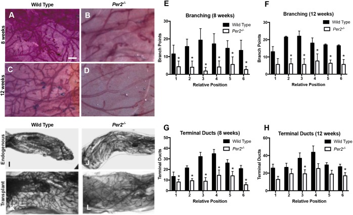
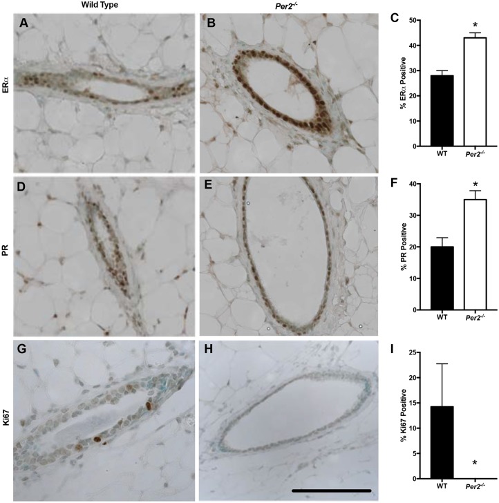
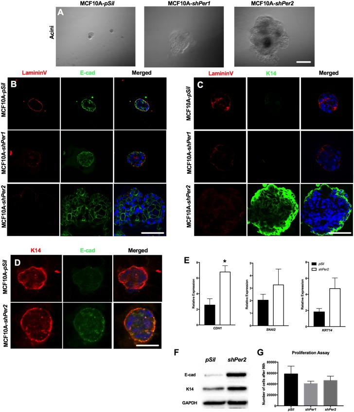
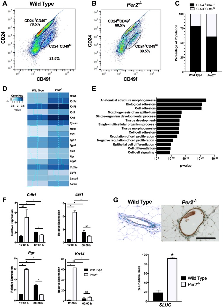
The molecular clock plays key roles in daily physiological functions, development and cancer. Period 2 (PER2) is a repressive element, which inhibits transcription activated by positive clock elements, resulting in diurnal cycling of genes. However, there are gaps in our understanding of the role of the clock in normal development outside of its time-keeping function. Here, we show that PER2 has a noncircadian function that is crucial to mammalian mammary gland development. Virgin Per2-deficient mice, Per2-/- , have underdeveloped glands, containing fewer bifurcations and terminal ducts than glands of wild-type mice. Using a transplantation model, we show that these changes are intrinsic to the gland and further identify changes in cell fate commitment. Per2-/- mouse mammary glands have a dual luminal/basal phenotypic character in cells of the ductal epithelium. We identified colocalization of E-cadherin and keratin 14 in luminal cells. Similar results were demonstrated using MCF10A and shPER2 MCF10A human cell lines. Collectively this study reveals a crucial noncircadian function of PER2 in mammalian mammary gland development, validates the Per2-/- model, and describes a potential role for PER2 in breast cancer.

Keywords: Mammary gland development; Molecular clock; PER2.


Conflict of interest statement

Competing interestsThe authors declare no competing or financial interests.

Figures

Fig. 1.
Fig. 1.
Disruption of branching morphogenesis in Per2−/− virgin mice. (A-D) Animal development stage is indicated along the left edge of panels. Representative images of normal branching and development of terminal ducts in a WT animal are shown (A,C). Representative images of Per2−/− mammary glands showing fewer bifurcations than in WT mammary glands (B,D). (E-H) The number of branch points (E,F) and terminal ducts (G,H) at the relative positions differed significantly between the WT and Per2−/− mice (*P<0.05; error bars indicate s.e.m.; n=3). (I-L) Mammary epithelia from WT and Per2−/− mice were transplanted contralaterally into the cleared fat pads of 21-day-old syngeneic mice and analyzed after 8 weeks of outgrowth. A phenotype consistent with Per2−/− mice was observed in the transplanted mutant glands. Per2−/−: n=4; WT: n=3. Scale bar: 500 μm.
Fig. 2.
Fig. 2.
Altered expression of luminal and basal markers in Per2−/− mammary glands. (A,B) H&E staining of mammary gland sections from adult WT, Per1−/− and Per2−/− mice. (C-F) IF staining for myoepithelial and luminal cell markers, SMA and MUC1 (C,D), K14/p63, K14/SMA, K14/E-cad and K14/K8 (E,F), showing inappropriate localization of myoepithelial and luminal markers. (G) Quantification of K14-positive cells in WT and Per2−/− virgin mice (*P<0.05; error bars indicate s.e.m.). (H) IF staining of mammary gland sections in WT and Per2−/− mice at lactation day 2 (L2), showing colocalization (arrows) of SMA/p63 expression (upper panels) and normal K14 expression (lower panels). Per2−/−: n=4; WT: n=3. Scale bars: 50 μm.
Fig. 3.
Fig. 3.
Altered hormone receptor status in Per2−/− virgin mice. (A-F) IHC staining indicated a significant increase in the number of ERα-positive (A-C) and PR-positive (D-F) cells in virgin Per2−/− mice compared with WT mice. (G-I) Staining for Ki67 revealed ∼15% Ki67-positive cells in WT mice, but almost no Ki67-positive cells in 10-week-old virgin Per2−/− mice. *P<0.05; error bars indicate s.e.m. Per2−/−: n=4; WT: n=3. Scale bar: 50 μm.
Fig. 4.
Fig. 4.
MCF10A (pSIL, shPER1, shPER2) human MECs in 3D culture. (A) MCF10A normal (pSIL) and shPER1 cells formed hollow spherical structures, indicating proper acini formation. MCF10A-shPER2 cells developed large, multilayered and filled in spherical structures, inconsistent with proper development. (B) Staining of a luminal marker (E-cad) indicates proper development in pSIL cells and shPER1 cells, yet an increase in the amount of E-cad fluorescence in shPER2 cells. (C) Staining of basal markers (lamininV and K14) indicates proper development in pSIL cells and shPER1 cells, yet improper localization in shPER2 cells. (D) K14 and E-cad staining showing colocalization in shPER2 acini. (E) qPCR analysis of luminal and basal markers indicated that CDH1, SNAI2 and KRT14 levels relative to β-actin were higher in the PER2 knockdown cells than in normal cells. (F) KRT14 and E-cad protein levels were also higher in the PER2 knockdown cells than in normal cells. (G) Proliferation assay results of MCF10A pSIL, shPER1 and shPER2 cell lines following 96 h under normal culture conditions. *P<0.05; error bars indicate s.e.m. Scale bars: 50 μm.
Fig. 5.
Fig. 5.
Cell lineage and RNA-Seq analysis of WT and Per2−/− MECs. (A-C) Per2−/− MECs showed a 19.55% reduction in luminal progenitors (CD24hi) and 6.63% increase in myoepithelial/basal-like cells (CD49fhi) when compared with WT cells. (D) RNA-Seq results of WT and Per2−/− mice, showing that genes associated with epithelial and basal phenotypes, including Krt14, Krtr5, Krt18 and Krt8, were upregulated in Per2−/− MECs. (E) GOrilla was used to identify biological pathways differentially regulated between the WT and Per2−/− mice. (F) qPCR analysis of luminal and basal markers confirmed that Chd1, Esr1, Pgr and Krt14 levels were higher relative to 18 s in Per2−/− MECs compared with WT MECs at 12:00 h. At 00:00 h, Chd1 and Pgr levels were lower in Per2−/− MECs compared with WT MECs, but no differences in the levels of Esr1 and Krt14 were observed. (G) SLUG staining showed a significant increase in SLUG in Per2−/− mouse mammary tissues compared with WT tissues. *P<0.05; error bars indicate s.e.m. Per2−/−: n=13; WT: n=10. Scale bar: 50 μm.

References

    1. Alvarez J. D., Chen D., Storer E. and Sehgal A. (2003). Non-cyclic and developmental stage-specific expression of circadian clock proteins during murine spermatogenesis. Biol. Reprod. 69, 81-91. 10.1095/biolreprod.102.011833 - DOI - PubMed
    1. Asselin-Labat M.-L., Sutherland K. D., Barker H., Thomas R., Shackleton M., Forrest N. C., Hartley L., Robb L., Grosveld F. G., van der Wees J. et al. (2007). Gata-3 is an essential regulator of mammary-gland morphogenesis and luminal-cell differentiation. Nat. Cell Biol. 9, 201-209. 10.1038/ncb1530 - DOI - PubMed
    1. Bae K., Jin X., Maywood E. S., Hastings M. H., Reppert S. M. and Weaver D. R. (2001). Differential functions of mPer1, mPer2, and mPer3 in the SCN circadian clock. Neuron 30, 525-536. 10.1016/S0896-6273(01)00302-6 - DOI - PubMed
    1. Bolos V., Peinado H., Pérez-Moreno M. A., Fraga M. F., Esteller M. and Cano A. (2003). The transcription factor Slug represses E-cadherin expression and induces epithelial to mesenchymal transitions: a comparison with Snail and E47 repressors. J. Cell Sci. 116, 499-511. 10.1242/jcs.00224 - DOI - PubMed
    1. Borgs L., Beukelaers P., Vandenbosch R., Nguyen L., Moonen G., Maquet P., Albrecht U., Belachew S. and Malgrange B. (2009). Period 2 regulates neural stem/progenitor cell proliferation in the adult hippocampus. BMC Neurosci. 10, 30 10.1186/1471-2202-10-30 - DOI - PMC - PubMed

Publication types