Review
. 2018 Feb 28;9(3):330.
doi: 10.1038/s41419-017-0023-6.

Mitofusin 2: from functions to disease

Affiliations
Review

Mitofusin 2: from functions to disease

Riccardo Filadi et al. Cell Death Dis. .

Abstract

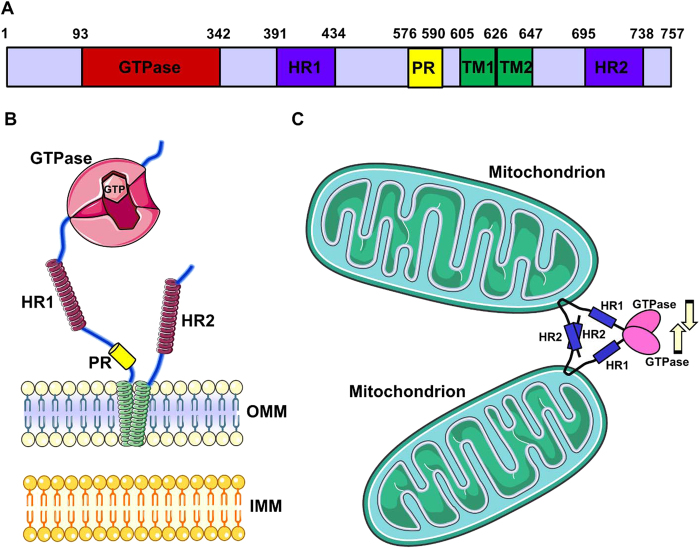
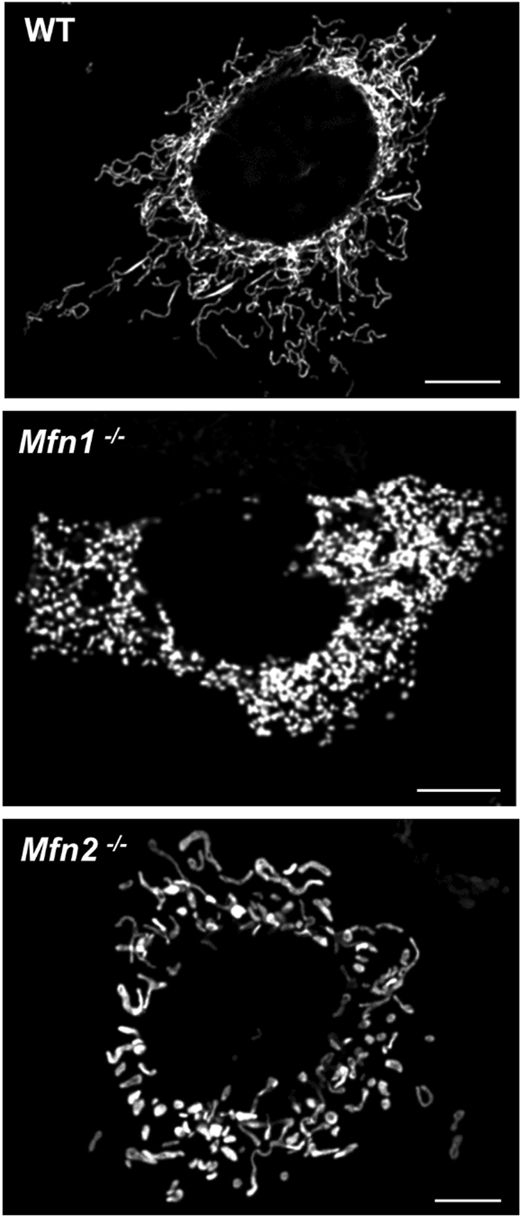
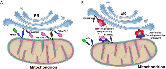
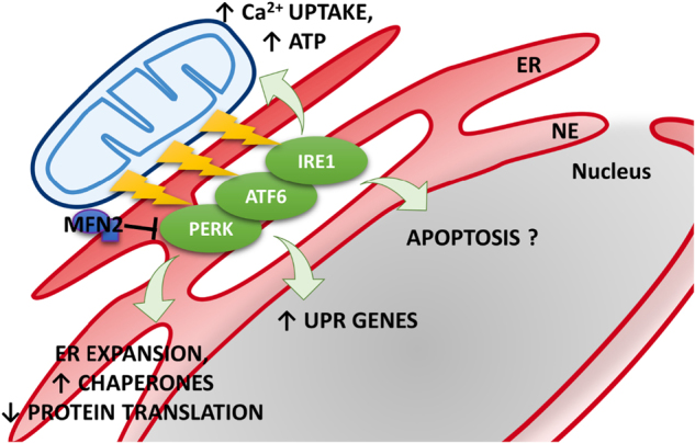
Mitochondria are highly dynamic organelles whose functions are essential for cell viability. Within the cell, the mitochondrial network is continuously remodeled through the balance between fusion and fission events. Moreover, it dynamically contacts other organelles, particularly the endoplasmic reticulum, with which it enterprises an important functional relationship able to modulate several cellular pathways. Being mitochondria key bioenergetics organelles, they have to be transported to all the specific high-energy demanding sites within the cell and, when damaged, they have to be efficiently removed. Among other proteins, Mitofusin 2 represents a key player in all these mitochondrial activities (fusion, trafficking, turnover, contacts with other organelles), the balance of which results in the appropriate mitochondrial shape, function, and distribution within the cell. Here we review the structural and functional properties of Mitofusin 2, highlighting its crucial role in several cell pathways, as well as in the pathogenesis of neurodegenerative diseases, metabolic disorders, cardiomyopathies, and cancer.


Conflict of interest statement

The authors declare no competing financial interests

Figures

Fig. 1
Fig. 1. MFN2 structure
a The scheme represents the linear structure of MFN2. Note the large N-terminal GTPase domain, followed by the HR1-domain, the PR domain, the two TM domains, and the C-terminal HR2 domain. The numbers above indicate the initial and the terminal amino acids of the corresponding domains. b The cartoon represents MFN2 topology, with two very close TM domains crossing the OMM (green helices) and the indicated cytosolic portions. Note the GTPase domain with two GTP-binding pockets. c Scheme of the OMM-fusion activity of MFNs. Tethering is mediated by the interaction between HR2 domains belonging to MFNs on opposite OMM. Recent data suggest that dimerization of the GTPase domains, as well as a power stroke due to GTP hydrolysis, may be important for fusion (see text for details)
Fig. 2
Fig. 2. Effects of MFNs ablation on mitochondrial morphology
Representative confocal microscopy images of WT, Mfn1−/− and Mfn2−/− MEF cells, expressing a mitochondrial matrix-targeted RFP. Scale bar: 5 μm. Note the fragmented mitochondrial network in MFNs ablated cells, with creation of small spheres in Mfn1−/− MEFs and of more enlarged structures of variable size in Mfn2−/− MEFs
Fig. 3
Fig. 3. Alternative models for MFN2-mediated ER−mitochondria tethering
a The scheme represents the classical view of MFN2 as a positive modulator of ER−mitochondria juxtaposition. In this model, MFN2 on ER membrane engages MFN2 or MFN1 on OMM, mediating the tethering between the two organelles. b The scheme represents the model of MFN2 as a negative modulator of ER−mitochondria juxtaposition. According to this view, MFN2 on both the ER and the OMM interacts with and sequesters still unknown tethering subunits (left), hindering their assembling into a functional tethering complex (represented on the right)
Fig. 4
Fig. 4. ER stress induced by MFN2 depletion
Mfn2 ablation induces the activation of UPR proteins located in ER membranes. In particular, MFN2 has been proposed as an upstream modulator of PERK that under basal conditions maintains the kinase inactive. Once activated, UPR works by expanding the ER, upregulating chaperones and inhibiting protein translation. Mitochondria display higher Ca2+ uptake and ATP production to support the higher energy demand. It is still unclear whether Mfn2 ablation induces an increase or decrease of ER stress-induced apoptosis
Fig. 5
Fig. 5. Schematic representation of MFN2 with its most common mutations linked to CMTA2
The scheme represents MFN2 protein and depicts amino acid mutations associated to CMT2A. The color refers to the domain affected: red, GTPase domain; blue, HR1/2; green, TM domains; black, linker regions. In bold, the more frequent mutations involving the arginine (R) in position 94

References

    1. Friedman JR, et al. ER tubules mark sites of mitochondrial division. Science. 2011;334:358–362. doi: 10.1126/science.1207385. - DOI - PMC - PubMed
    1. Gomes LC, Di Benedetto G, Scorrano L. During autophagy mitochondria elongate, are spared from degradation and sustain cell viability. Nat. Cell. Biol. 2011;13:589–598. doi: 10.1038/ncb2220. - DOI - PMC - PubMed
    1. Rambold AS, Kostelecky B, Elia N, Lippincott-Schwartz J. Tubular network formation protects mitochondria from autophagosomal degradation during nutrient starvation. Proc. Natl. Acad. Sci. USA. 2011;108:10190–10195. doi: 10.1073/pnas.1107402108. - DOI - PMC - PubMed
    1. Pendin D, Filadi R, Pizzo P. The concerted action of mitochondrial dynamics and positioning: new characters in cancer onset and progression. Front. Oncol. 2017;7:102. doi: 10.3389/fonc.2017.00102. - DOI - PMC - PubMed
    1. Smirnova E, Griparic L, Shurland DL, van der Bliek AM. Dynamin-related protein Drp1 is required for mitochondrial division in mammalian cells. Mol. Biol. Cell. 2001;12:2245–2256. doi: 10.1091/mbc.12.8.2245. - DOI - PMC - PubMed

Publication types

MeSH terms

LinkOut - more resources