Review
. 2018 Feb 28;9(3):335.
doi: 10.1038/s41419-017-0215-0.

A key role for MAM in mediating mitochondrial dysfunction in Alzheimer disease

Affiliations
Review

A key role for MAM in mediating mitochondrial dysfunction in Alzheimer disease

Estela Area-Gomez et al. Cell Death Dis. .

Abstract

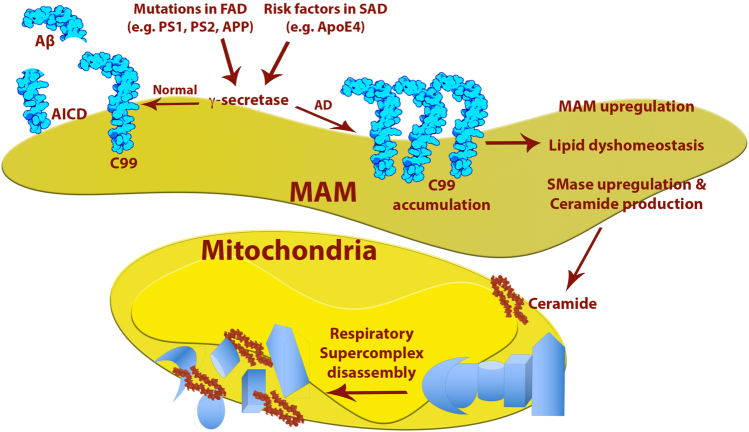
In the last few years, increased emphasis has been devoted to understanding the contribution of mitochondria-associated endoplasmic reticulum (ER) membranes (MAM) to human pathology in general, and neurodegenerative diseases in particular. A major reason for this is the central role that this subdomain of the ER plays in metabolic regulation and in mitochondrial biology. As such, aberrant MAM function may help explain the seemingly unrelated metabolic abnormalities often seen in neurodegeneration. In the specific case of Alzheimer disease (AD), besides perturbations in calcium and lipid homeostasis, there are numerous documented alterations in mitochondrial behavior and function, including reduced respiratory chain activity and oxidative phosphorylation, increased free radical production, and altered organellar morphology, dynamics, and positioning (especially perinuclear mitochondria). However, whether these alterations are primary events causative of the disease, or are secondary downstream events that are the result of some other, more fundamental problem, is still unclear. In support of the former possibility, we recently reported that C99, the C-terminal processing product of the amyloid precursor protein (APP) derived from its cleavage by β-secretase, is present in MAM, that its level is increased in AD, and that this increase reduces mitochondrial respiration, likely via a C99-induced alteration in cellular sphingolipid homeostasis. Thus, the metabolic disturbances seen in AD likely arise from increased ER-mitochondrial communication that is driven by an increase in the levels of C99 at the MAM.


Conflict of interest statement

The authors declare that they have no conflict of interest.

Figures

Fig. 1
Fig. 1
a Representative mitochondrial morphology in AD-mutant cells. Human fibroblasts were stained with MitoTracker (red) and anti-tubulin (green). Note the relatively dispersed distribution of the mitochondria in the control, whereas they are more perinuclear in the FAD-PS1M146L and FAD-PS1A246E cells. b MEFs in which PS1 was knocked down (by small hairpin RNA). Cells were stained as in a. Note relatively dispersed distribution of the mitochondria in the control, whereas they are more perinuclear in the PS1-knockdown cells. This phenotype could be rescued by overexpression of WT human PS1, but not by expression of a human pathogenic PS1 mutation (A246E)
Fig. 2
Fig. 2. Immunohistochemistry to detect mitochondria (FeS subunit of complex III of the mitochondrial respiratory chain) in the hippocampus (CA1 region) from a FAD patient with a PS1 mutation (A434C).
(Upper panels) Control subject (left), with four indicated neurons (ad) magnified at right. Note relatively uniform stain (brown), indicating a homogeneous distribution of mitochondria in the cell body. (Lower panels) FAD-PS1A434C patient. Notation as in upper panel. Note the perinuclear distribution of immunostain (brown rings; arrowheads), with a relative paucity of immunostain in the distal regions of the cell body (asterisks)
Fig. 3
Fig. 3. Model of the “MAM hypothesis” for the pathogenesis of AD, with emphasis on the mitochondrial alterations.
See text for details

References

    1. Alzheimer’s Association. Alzheimer’s disease facts and figures. Alzheimers Dement. 2011;7:208–244. doi: 10.1016/j.jalz.2011.02.004. - DOI - PubMed
    1. Goedert M, Spillantini MG. A century of Alzheimer’s disease. Science. 2006;314:777–781. doi: 10.1126/science.1132814. - DOI - PubMed
    1. Hardy JA, Higgins GA. Alzheimer’s disease: the amyloid cascade hypothesis. Science. 1992;256:184–185. doi: 10.1126/science.1566067. - DOI - PubMed
    1. Nunomura A, et al. Oxidative damage is the earliest event in Alzheimer disease. J. Neuropathol. Exp. Neurol. 2001;60:759–767. doi: 10.1093/jnen/60.8.759. - DOI - PubMed
    1. Manczak M, Sheiko T, Craigen WJ, Reddy PH. Reduced VDAC1 protects against Alzheimer’s disease, mitochondria, and synaptic deficiencies. J. Alzheimers Dis. 2013;37:679–690. - PMC - PubMed

Publication types

MeSH terms

Substances