Review
. 2018 Feb 28;9(3):328.
doi: 10.1038/s41419-017-0025-4.

Mitochondria-associated ER membranes (MAMs) and lysosomal storage diseases

Affiliations
Review

Mitochondria-associated ER membranes (MAMs) and lysosomal storage diseases

Ida Annunziata et al. Cell Death Dis. .

Abstract

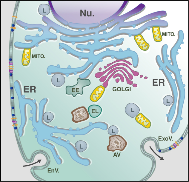
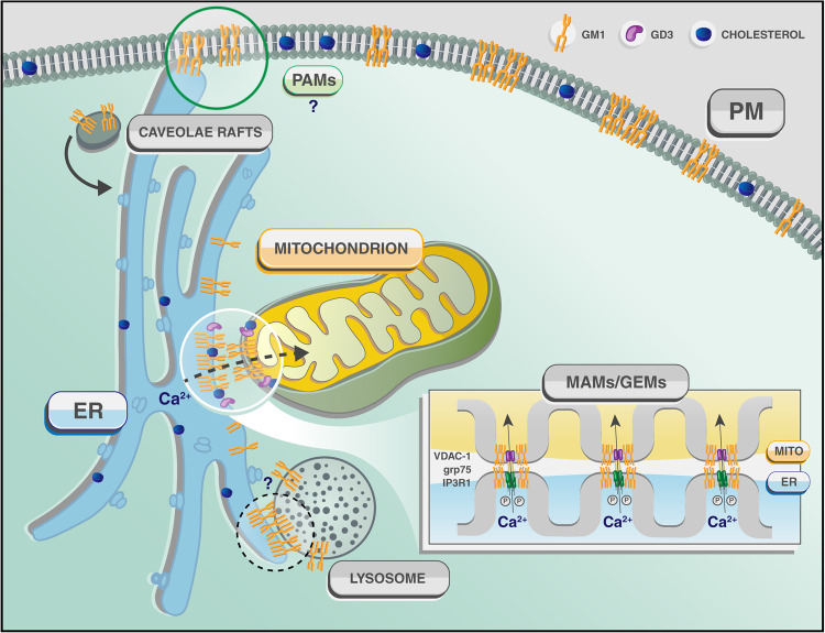
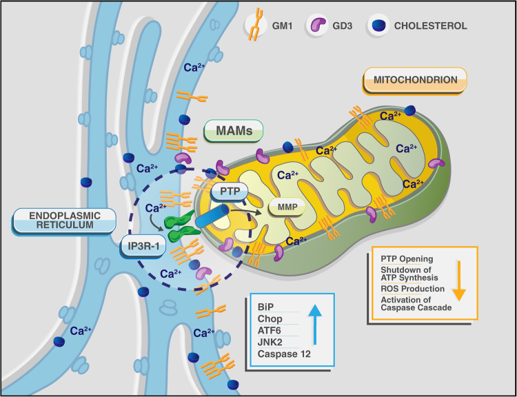
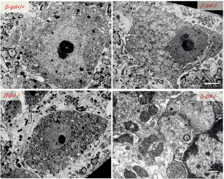
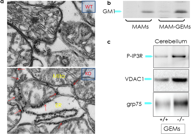
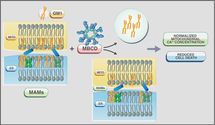
Lysosomal storage diseases (LSDs) comprise a large group of disorders of catabolism, mostly due to deficiency of a single glycan-cleaving hydrolase. The consequent endo-lysosomal accumulation of undigested or partially digested substrates in cells of virtually all organs, including the nervous system, is diagnostic of these diseases and underlies pathogenesis. A subgroup of LSDs, the glycosphingolipidoses, are caused by deficiency of glycosidases that process/degrade sphingolipids and glycosphingolipids (GSLs). GSLs are among the lipid constituents of mammalian membranes, where they orderly distribute and, together with a plethora of membrane proteins, contribute to the formation of discrete membrane microdomains or lipid rafts. The composition of intracellular membranes enclosing organelles reflects that at the plasma membrane (PM). Organelles have the tendencies to tether to one another and to the PM at specific membrane contact sites that, owing to their lipid and protein content, resemble PM lipid rafts. The focus of this review is on the MAMs, mitochondria associated ER membranes, sites of juxtaposition between ER and mitochondria that function as biological hubs for the exchange of molecules and ions, and control the functional status of the reciprocal organelles. We will focus on the lipid components of the MAMs, and highlight how failure to digest or process the sialylated GSL, GM1 ganglioside, in lysosomes alters the lipid conformation and functional properties of the MAMs and leads to neuronal cell death and neurodegeneration.


Conflict of interest statement

The authors declare no competing financial interest.

Figures

Fig. 1
Fig. 1. Membrane contact sites in Eukaryotes
Schematic representation of an eukaryotic cell and its interorganellar membrane contact sites. The vast network of the ER participates in multiple membrane contact sites with the membranes of mitochondria (MITO.), PM, early endosome (EE), lysosome (L) and Golgi. Additionally, lysosomes can tether with mitochondrial and nuclear (Nu.) membranes. EL endolysosomes; AV autophagyc vacuoles; EnV endocytic vesicles; ExoV exocytic vesicles
Fig. 2
Fig. 2
Schematic rendering of the contact sites between ER and mitochondria (MAMs), ER and PM (PAMs) and potentially ER and lysosomes (?) that explain the redistribution and buildup of GM1 in the ER membranes and the consequent activation of the apoptotic process leading to neuronal cell death in β-gal−/− mice
Fig. 3
Fig. 3. Schematic representation of a single MAM, depicted as a functional hub for the aberrant transfer of Ca2+ between ER and mitochondria, leading to ER- and mitochondria- mediated neuronal cell death in GM1-gangliosidosis
The figure also lists the principal effectors of the apoptotic process described in Tessitore et al., 2004 and Sano et al., 2009
Fig. 4
Fig. 4. Ultrastructural abnormalities in the CNS of β-Gal−/− mice
Transmission electron microscopy of spinal cord neurons from 3-month-old β-Gal−/− and β-Gal+/+ mice shows evidence in the affected mouse of an expanded lysosomal compartment with enlarged lysosomes filled with membranous material due to accumulation of GM1-ganglioside. Scale bars: 1 μm; lover right panel 0.5 μm. Adapted from the original article Tessitore et al., 2004 with the permission of Elsevier
Fig. 5
Fig. 5. GM1 accumulation in the GEMs alters MAMs dynamics
a Representative electron micrographs of mitochondria isolated from β-Gal+/+ and β-Gal−/− brains showed larger areas of juxtaposition between ER and mitochondria in the β-Gal−/− preparations compared to the WT. b TLC analysis of lipids from the purified MAMs, and the Triton-extracted (Triton extr. MAMs) and Triton-insoluble fractions (GEMs) of the MAMs demonstrated the buildup of GM1 in all β-Gal−/− fractions. c Increased levels of phosphorylated IP3R1, VDAC1, and GRP75 were detected in the GEMs extracted from β-Gal−/− brains compared to β-Gal+/+ brains. Adapted from the original article Sano et al., 2009 with the permission of Elsevier
Fig. 6
Fig. 6. Schematic rendering of the effects of MBCD on MAMs/GEMs in GM1-accumulating cells
MBCD efficiently extracts GM1 from these microdomains and, in turn, reverts mitochondrial Ca2+ overload and apoptosis

References

    1. Ji CH, Kwon YT. Crosstalk and Interplay between the ubiquitin-proteasome system and autophagy. Mol. Cell. 2017;40:441–449. - PMC - PubMed
    1. Saftig, P. Physiology of the lysosome. In: Mehta, A., Beck, M., Sunder-Plassmann, G. (eds). Fabry Disease: Perspectives from 5 Years of FOS Oxford PharmaGenesis; Oxford, (2006) Chapter 3. - PubMed
    1. Saftig P, Klumperman J. Lysosome biogenesis and lysosomal membrane proteins: trafficking meets function. Nat. Rev. Mol. Cell. Biol. 2009;10:623–635. doi: 10.1038/nrm2745. - DOI - PubMed
    1. Carette JE, Raaben M, Wong AC, Herbert AS, Obernosterer G, Mulherkar N, et al. Ebola virus entry requires the cholesterol transporter Niemann-Pick C1. Nature. 2011;477:340–343. doi: 10.1038/nature10348. - DOI - PMC - PubMed
    1. Ndungo, E., Herbert, A. S., Raaben, M., Obernosterer, G., Biswas, R., Miller, E. H. et al. A single residue in ebola virus receptor npc1 influences cellular host range in reptiles. mSphere1 e00007-16 (2016) - PMC - PubMed

Publication types

MeSH terms

LinkOut - more resources