. 2018 Feb 28;9(1):863.
doi: 10.1038/s41467-018-03318-5.

FcαRI co-stimulation converts human intestinal CD103+ dendritic cells into pro-inflammatory cells through glycolytic reprogramming

Affiliations

FcαRI co-stimulation converts human intestinal CD103+ dendritic cells into pro-inflammatory cells through glycolytic reprogramming

Ivo S Hansen et al. Nat Commun. .

Abstract

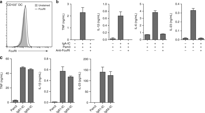
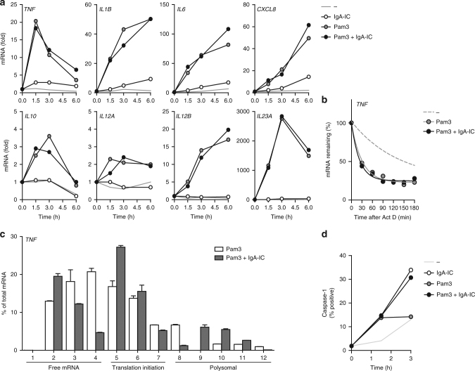
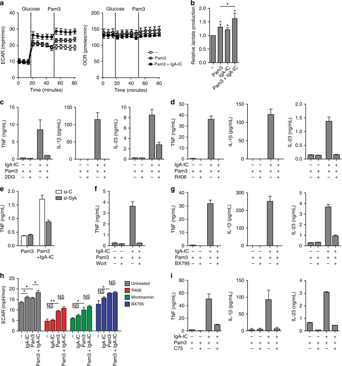
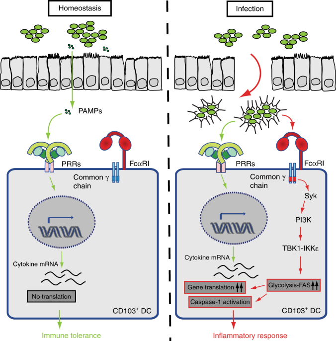
CD103+ dendritic cells (DC) are crucial for regulation of intestinal tolerance in humans. However, upon infection of the lamina propria this tolerogenic response is converted to an inflammatory response. Here we show that immunoglobulin A (IgA) immune complexes (IgA-IC), which are present after bacterial infection of the lamina propria, are important for the induction of inflammation by the human CD103+SIRPα+ DC subset. IgA-IC, by recognition through FcαRI, selectively amplify the production of proinflammatory cytokines TNF, IL-1β and IL-23 by human CD103+ DCs. These cells then enhance inflammation by promoting Th17 responses and activating human intestinal innate lymphoid cells 3. Moreover, FcαRI-induced cytokine production is orchestrated via upregulation of cytokine translation and caspase-1 activation, which is dependent on glycolytic reprogramming mediated by kinases Syk, PI3K and TBK1-IKKε. Our data suggest that the formation of IgA-IC in the human intestine provides an environmental cue for the conversion of a tolerogenic to an inflammatory response.


Conflict of interest statement

D.L.P.B. is a part-time employee of UCB. G.R.v.d.B. is currently employed by GlaxoSmithKline. The remaining authors declare no competing interests.

Figures

Fig. 1
Fig. 1
IgA opsonization of bacteria enhances production of proinflammatory cytokines by CD103+ DCs. CD103+ dendritic cells (DC) or moDCs were stimulated with 10 MOI S. aureus, which were untreated or opsonized with 5 mg/mL serum IgA for 1 h and washed. Experiments were performed in triplicate. After 24 h cytokine levels were analysed using ELISA, mean + s.e.m. Representative example of three experiments with different donors
Fig. 2
Fig. 2
IgA-IC promote proinflammatory cytokine production through synergy with various PRRs. a CD103+ dendritic cells (DC) were stimulated with Pam3CSK4 (Pam3), IgA immune complexes (IgA-IC), or a combination. b Cytokine production by CD103+ DCs stimulated with Pam3CSK4 or Pam3CSK4 combined with IgA-IC. Each pair of dots represents one donor. *p < 0.05, **p < 0.01, ***p < 0.001, Mann–Whitney U-test. c Cytokine production by moDCs stimulated with Pam3CSK4, IgA-IC, or a combination. d CD103+ DCs isolated from surgically resected intestine (1× ileum in grey, 3× colon in black) were stimulated with Pam3CSK4 or Pam3CSK4 combined with IgA-IC. Each pair of dots represents one donor. *p < 0.05. Student’s t-test. e Cytokine production by CD103+ DCs stimulated with different pathogen-associated molecular patterns (PAMP) alone or combined with IgA-IC. Stimulated receptors were TLR2 (LTA), TLR4 (LPS), TLR3 (Poly IC), TLR7/8 (CLO97), TLR2/NOD2 (PGN), NOD2 (MDP), and Dectin-1 (Curdlan). Experiments were performed in triplicate. After 24 h cytokine levels were analysed using ELISA, mean + s.e.m. Representative example of five (a) or three (c and e) experiments using different donors
Fig. 3
Fig. 3
Complexed IgA1 and IgA2 amplify proinflammatory cytokine production through FcαRI. a FcαRI expression on unstimulated CD103+ dendritic cells (DC) was analysed using flow cytometry. Light grey histogram indicates background staining. b Prior to stimulation, CD103+ DCs were incubated with a blocking antibody against FcαRI and stimulated with Pam3CSK4 (Pam3) alone or Pam3CSK4 together with IgA immune complexes (IgA-IC). c Cytokine production by CD103+ DCs stimulated with Pam3CSK4 alone or in combination with IgA1-IC or IgA2-IC. Experiments b and c were performed in triplicate. After 24 h cytokine levels were analysed using ELISA, mean + s.e.m. Representative example (ac) of three experiments using different donors
Fig. 4
Fig. 4
Co-stimulation of CD103+ DCs with IgA-IC promotes Th17 responses and activates intestinal ILC3. a CD103+ dendritic cells (DC) were stimulated with Pam3CSK4 (Pam3) alone or Pam3CSK4 together with IgA immune complexes (IgA-IC) and co-cultured with CD4+ T Cells. After T-cell outgrowth, resting cells were re-stimulated and after 24 h supernatant was assayed with ELISA, mean + s.e.m. Experiments were performed in triplicate. b, c Supernatant from CD103+ DCs which were stimulated with Pam3CSK4 or Pam3CSK4 with IgA-IC was transferred onto human intestinal ILC3 and cultured for 7 days and analysed for mRNA expression (b) or protein expression (c). ND, not detected. Experiments were performed in triplicate. mRNA expression of indicated genes were assayed using qPCR (normalized to GAPDH expression) mean + s.e.m. Supernatant was analysed using ELISA, mean + s.e.m. Representative example of three (a, b) or two (c) experiments using different donors
Fig. 5
Fig. 5
FcαRI stimulation promotes cytokine gene translation and induces caspase-1 activation. a CD103+ dendritic cells (DC) were stimulated with Pam3CSK4 (Pam3), IgA immune complexes (IgA-IC), or a combination and analysed for mRNA expression of indicated genes using qPCR (normalized to GAPDH expression, fold increase compared to unstimulated control). b mRNA stability of TNF of CD103+ DCs which were unstimulated or treated with Pam3CSK4 or co-stimulation with IgA-IC was determined after addition of 10 µg/mL actinomycin D (Act D) to prevent de novo synthesis of mRNA. TNF expression was analysed using qPCR (normalized to GAPDH). c Lysates of CD103+ DCs stimulated for 3 h with Pam3CSK4 or Pam3CSK4 with IgA-IC were loaded on sucrose gradients to measure mRNA translation of TNF (normalized to GAPDH, qPCR was performed in duplicate, mean + s.e.m.). d Induction of caspase-1 activation in CD103+ DCs after stimulation with Pam3CSK4, IgA-IC, or a combination was measured using caspase-1 binding compound FAM-YVAD-FMK (FLICA) by flow cytometry. Representative example (ad) of three independent experiments
Fig. 6
Fig. 6
FcαRI-induced upregulation of cytokine production is dependent on glycolytic reprogramming through Syk, PI3K, and TBK1-IKKε. a Real-time changes in the extracellular acidification rate (ECAR) and oxygen consumption rate (OCR) of CD103+ dendritic cells (DC) left unstimulated (open circles), stimulated with Pam3CSK4 (Pam3) alone (grey circles), or Pam3CSK4 with IgA immune complexes (IgA-IC) (black circles). IgA-IC are present from the start of the experiment. The dotted line indicates initiation of further treatment. Experiments were performed in quadruplicate, mean + s.e.m. b Relative lactate production of CD103+ DCs after 24 h of treatment. Pooled data from five experiments. *p < 0.05. Student’s t test. c, d Cytokine production by CD103+ DCs stimulated with Pam3CSK4, IgA-IC, or a combination after treatment with 10 mM glycolysis inhibitor 2-deoxy-D-glucose (2DG) (c) or 1 µM of syk inhibitor R406 (d). e TNF production by CD103+ DCs silenced using specific si-RNA for Syk (si-Syk) or non-targeted control (si-C). f, g Cytokine production by CD103+ DCs stimulated with Pam3CSK4, IgA-IC, or a combination after treatment with 100 nM PI3K inhibitor Wortmannin (Wort) (f) or 1 µM TBK1-IKKε inhibitor BX795 (g). h Differences in ECAR levels of CD103+ DCs stimulated with Pam3CSK4, IgA-IC or a combination in the presence of inhibitors for Syk (R406), PI3K (Wort), or TBK1-IKKε (BX795). Experiments were performed in quadruplicate, mean + s.e.m. *p < 0.05, **p < 0.01, NS not significant. Student’s t test. i Cytokine production by CD103+ DCs stimulated with Pam3CSK4, IgA-IC, or a combination after treatment with 20 µM fatty acid synthase inhibitor C75. Experiments cg, i were performed in triplicate. After 24 h cytokine levels were analysed using ELISA, mean + s.e.m. Representative example (a, ci) of three independent experiments
Fig. 7
Fig. 7
Model for inflammatory response upon FcαRI-TLR cross-talk. Under homeostatic conditions CD103+ dendritic cells (DC) are activated by pathogen-associated molecular patterns (PAMP) through leakage or sampling of the lumen. This induces inflammatory cytokine mRNA transcription, but no protein production. Upon infection, IgA immune complexes (IgA-IC) provide a second signal through FcαRI triggering Syk, PI3K, and TBK1-IKKε activation, which increases the glycolytic flux and fatty acid synthesis (FAS) that results in an inflammatory response mediated by increased mRNA translation and caspase-1 activation

References

    1. Bekiaris V, Persson EK, Agace WW. Intestinal dendritic cells in the regulation of mucosal immunity. Immunol. Rev. 2014;260:86–101. doi: 10.1111/imr.12194. - DOI - PubMed
    1. Farache J, Zigmond E, Shakhar G, Jung S. Contributions of dendritic cells and macrophages to intestinal homeostasis and immune defense. Immunol. Cell. Biol. 2013;91:232–239. doi: 10.1038/icb.2012.79. - DOI - PubMed
    1. Scott CL, Aumeunier AM, Mowat AM. Intestinal CD103+ dendritic cells: master regulators of tolerance? Trends Immunol. 2011;32:412–419. doi: 10.1016/j.it.2011.06.003. - DOI - PubMed
    1. Johansson-Lindbom B, et al. Functional specialization of gut CD103+ dendritic cells in the regulation of tissue-selective T cell homing. J. Exp. Med. 2005;202:1063–1073. doi: 10.1084/jem.20051100. - DOI - PMC - PubMed
    1. Schulz O, et al. Intestinal CD103+, but not CX3CR1+, antigen sampling cells migrate in lymph and serve classical dendritic cell functions. J. Exp. Med. 2009;206:3101–3114. doi: 10.1084/jem.20091925. - DOI - PMC - PubMed

Publication types