

Keratinizing pleomorphic adenoma: An unusual case report
- PMID: 29491610
- PMCID: PMC5824522
- DOI: 10.4103/jomfp.JOMFP_200_17
Keratinizing pleomorphic adenoma: An unusual case report
Abstract
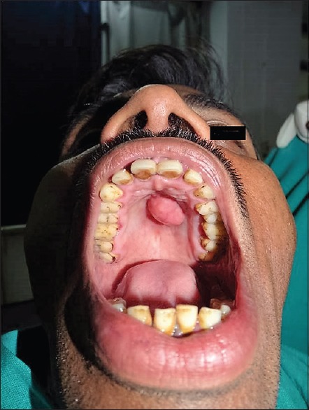
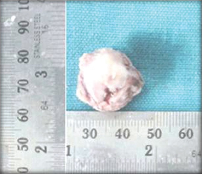
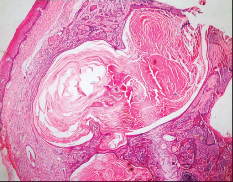
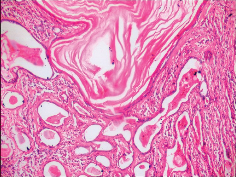
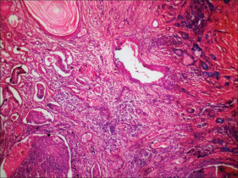
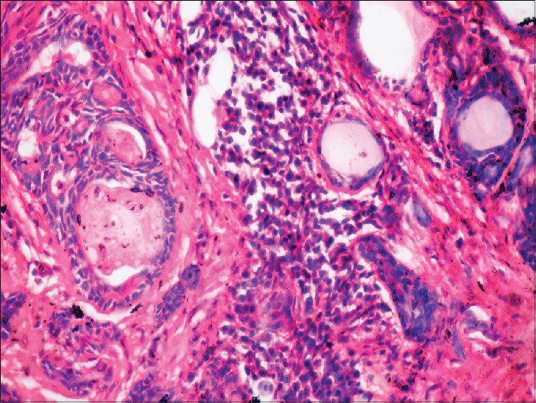
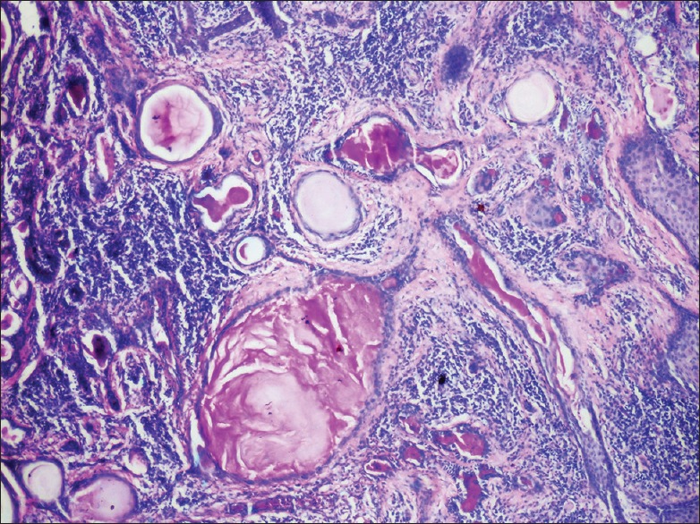
Pleomorphic adenoma (PA) is the most common benign tumor of major or minor salivary glands. PA exhibits a great histological diversity, such as differentiation into oncocytic, sebaceous, mucinous, squamous, chondroid, osseous or adipose cells. Squamous metaplasia rarely results in the formation of extensive keratin-filled cyst lined by squamous epithelium. Extensive squamous metaplasia can be mistaken for malignancy, including mucoepidermoid carcinoma and squamous cell carcinoma. Here, we report a case of slowly enlarging PA with extensive squamous metaplasia and keratin cyst formations in a minor salivary gland in hard palate and discuss its microscopic features.
Keywords: Keratin; pleomorphic adenoma; squamous metaplasia.
Conflict of interest statement
There are no conflicts of interest.
Figures
References
-
- Abdul HA, Mandakini BT, Azhar F. Palatal pleomorphic adenoma with florid squamous metaplasia: A potential diagnostic pitfall. J Evol Med Dent Sci. 2012;1:96–101.
-
- Friedrich RE, Li L, Knop J, Giese M, Schmelzle R. Pleomorphic adenoma of the salivary glands: Analysis of 94 patients. Anticancer Res. 2005;25:1703–5. - PubMed
-
- Eveson JW, Cawson RA. Salivary gland tumours. A review of 2410 cases with particular reference to histological types, site, age and sex distribution. J Pathol. 1985;146:51–8. - PubMed
-
- Eveson JW, Auclair P, Gnepp DR, El-Naggar AK. Tumours of the salivary glands. In: Barnes L, Eveson JW, Reichart P, Sidransky D, editors. World Health Organization Classification of Tumours: Pathology and Genetics of Head and Neck Tumours. Lyon: IARC Press; 2005. pp. 209–81.
Publication types
LinkOut - more resources
Full Text Sources
Other Literature Sources
