

Case Reports
doi: 10.4103/jomfp.JOMFP_280_17.
Trigeminal neuralgia induced by odontogenic keratocyst associated with impacted supernumerary teeth: A rare case report
Affiliations
- PMID: 29491624
- PMCID: PMC5824508
- DOI: 10.4103/jomfp.JOMFP_280_17
Item in Clipboard
Case Reports
Trigeminal neuralgia induced by odontogenic keratocyst associated with impacted supernumerary teeth: A rare case report
J Oral Maxillofac Pathol.
2018 Jan.
Abstract
Odontogenic keratocyst(OKC)is a cyst oftooth origin with an aggressive behavior including a high recurrence rate, it has been rechristened to keratocystic odontogenic tumor(KCOT) as it be the reflects its neoplastic nature. We report a case of KCOT in association with an impacted supernumerary tooth along with Trigeminal Neuralgia, that subsided by itself after removal of the cyst.
Keywords: Odontogenic Keratocyst; Supernumerary tooth; Trigeminal Neuralgia.
Conflict of interest statement
There are no conflicts of interest.
Figures

Clinical picture
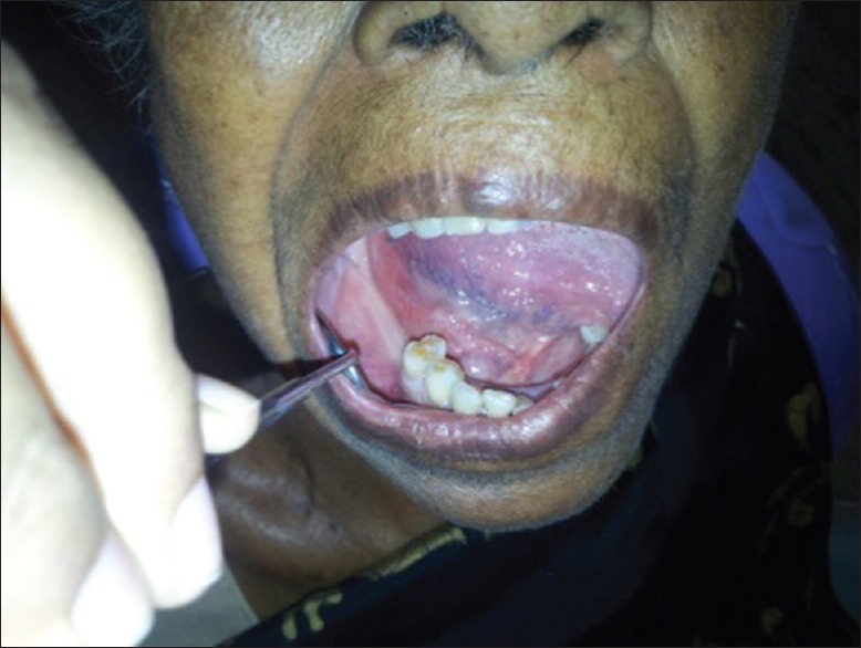
Intraoral examination revealed missing teeth in relation to 46, 47 and 48 with no buccal and lingual plate cortical expansion. Attrition and cervical abrasion present in relation to 44 and 45

Intraoral examination revealed missing teeth in relation to 46, 47 and 48 with no buccal and lingual plate cortical expansion. Attrition and cervical abrasion present in relation to 44 and 45

An orthopantomogram revealing a well-defined radiolucency in the right body of the mandible that surrounds impacted supernumerary teeth. Presence of 2 more supernumeraries in the maxillary 1st quadrant and 3rd quadrant.

Mandibular cross-section view

Panoramic view on cone-beam computed tomography

Axial view on cone-beam computed tomography

Cross-section view on cone-beam computed tomography

Soft-tissue incision placed distal to 45

A bony trough was placed on the alveolus

The lesion was retracted and here we can see the attachment of the lesion to the mandibular nerve

Complete removal of the lesion and the impacted tooth with thorough irrigation

Excised specimen

Sutures placed

H&E picture, odontogenic keratocyst
References
-
- Rapini RP, Bolognia JL, Jorizzo JL. Dermatology. 1, 2. St. Louis: Mosby; 2007. p. 101.
-
- Joffroy A, Levivier M, Massager N. Trigeminal neuralgia – Pathophysiology and treatment. Acta Neurol (Belg) 2001;101:20–5. - PubMed
-
- Scrivani SJ, Mathews ES, Maciewicz RJ. Trigeminal neuralgia. Oral Surg Oral Med Oral Pathol Oral Radiol Endod. 2005;100:527–38. - PubMed
-
- Adams CB. Trigeminal neuralgia: Pathogenesis and treatment. Br J Neurosurg. 1997;11:493–5. - PubMed
-
- Love S, Coakham HB. Trigeminal neuralgia: Pathology and pathogenesis. Brain. 2001;124:2347–60. - PubMed
Publication types
LinkOut - more resources
Full Text Sources
Other Literature Sources
