Case Reports
. 2018 Jan-Mar;13(1):157-160.
doi: 10.4103/1793-5482.181139.

Primary Giant Sphenotemporal Intradiploic Meningioma

Affiliations
Case Reports

Primary Giant Sphenotemporal Intradiploic Meningioma

Dipanker Singh Mankotia et al. Asian J Neurosurg. 2018 Jan-Mar.

Abstract

Intradiploic meningioma is a rare subset of meningioma accounting for 1% of all cases. Authors report a rare case of giant sphenotemporal intradiploic meningioma with orbital extension in a 27-year-old female. It was managed successfully with complete surgical excision and bony reconstruction using autologous split thickness bone graft.

Keywords: Intradiploic; intraosseous; meningioma; primary extradural meningioma; sphenotemporal.


Conflict of interest statement

There are no conflicts of interest.

Figures

Figure 1
Figure 1
Contrast-enhanced computed tomography head with bone window (a-c) demonstrating well-defined, lytic, sharply marginated intradiploic lesion marked with an arrow in the left sphenotemporal region reaching up to orbit and sphenoid sinus medially. Bony shell (arrow) is seen all around the lesion
Figure 2
Figure 2
Contrast magnetic resonance imaging demonstrating homogenously enhancing left sphenotemporal lesion in axial (a), sagittal (b), and coronal planes (c)
Figure 3
Figure 3
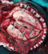
Intraoperative image demonstrating the pure extradural nature of lesion with intact dura
Figure 4
Figure 4
Post bony reconstruction using autologous split calvarial bone graft and titanium plates
Figure 5
Figure 5
Resected tumor tissue
Figure 6
Figure 6
Photomicrographs showing sheets of tumor cells with interspersed collagen bundles (a: H and E, ×40); polygonal cells with clear to eosinophilic cytoplasm (b: H and E, ×400); immunoreactivity for epithelial membrane antigen (c: Immunohistochemistry, ×400) and vimentin (d: Immunohistochemistry, ×400); immunonegative for cytokeratin (e: Immunohistochemistry, ×400) and CD34 (f: Immunohistochemistry: ×400)

References

    1. Lang FF, Macdonald OK, Fuller GN, DeMonte F. Primary extradural meningiomas: A report on nine cases and review of the literature from the era of computerized tomography scanning. J Neurosurg. 2000;93:940–50. - PubMed
    1. Liu Y, Wang H, Shao H, Wang C. Primary extradural meningiomas in head: A report of 19 cases and review of literature. Int J Clin Exp Pathol. 2015;8:5624–32. - PMC - PubMed
    1. Oka K, Hirakawa K, Yoshida S, Tomonaga M. Primary calvarial meningiomas. Surg Neurol. 1989;32:304–10. - PubMed
    1. Verma SK, Satyarthee G, Borkar SA, Singh M, Sharma BS. Orbital roof intradiploic meningioma in a 16-year-old girl. J Pediatr Neurosci. 2015;10:51–4. - PMC - PubMed
    1. Crawford TS, Kleinschmidt-DeMasters BK, Lillehei KO. Primary intraosseous meningioma. Case report. J Neurosurg. 1995;83:912–5. - PubMed

Publication types