. 2018 Oct;43(10):2702-2711.
doi: 10.1007/s00261-018-1519-y.

Computed tomography findings after radiofrequency ablation in locally advanced pancreatic cancer

Affiliations

Computed tomography findings after radiofrequency ablation in locally advanced pancreatic cancer

Steffi J E Rombouts et al. Abdom Radiol (NY). 2018 Oct.

Abstract

Purpose: The purpose of the study was to provide a systematic evaluation of the computed tomography(CT) findings after radiofrequency ablation (RFA) in locally advanced pancreatic cancer(LAPC).

Methods: Eighteen patients with intra-operative RFA-treated LAPC were included in a prospective case series. All CT-scans performed prior to RFA and 1 week and 3 months of post-RFA, according to standard regimen, were assessed by two radiologists in consensus, using standardized radiological scoring lists.

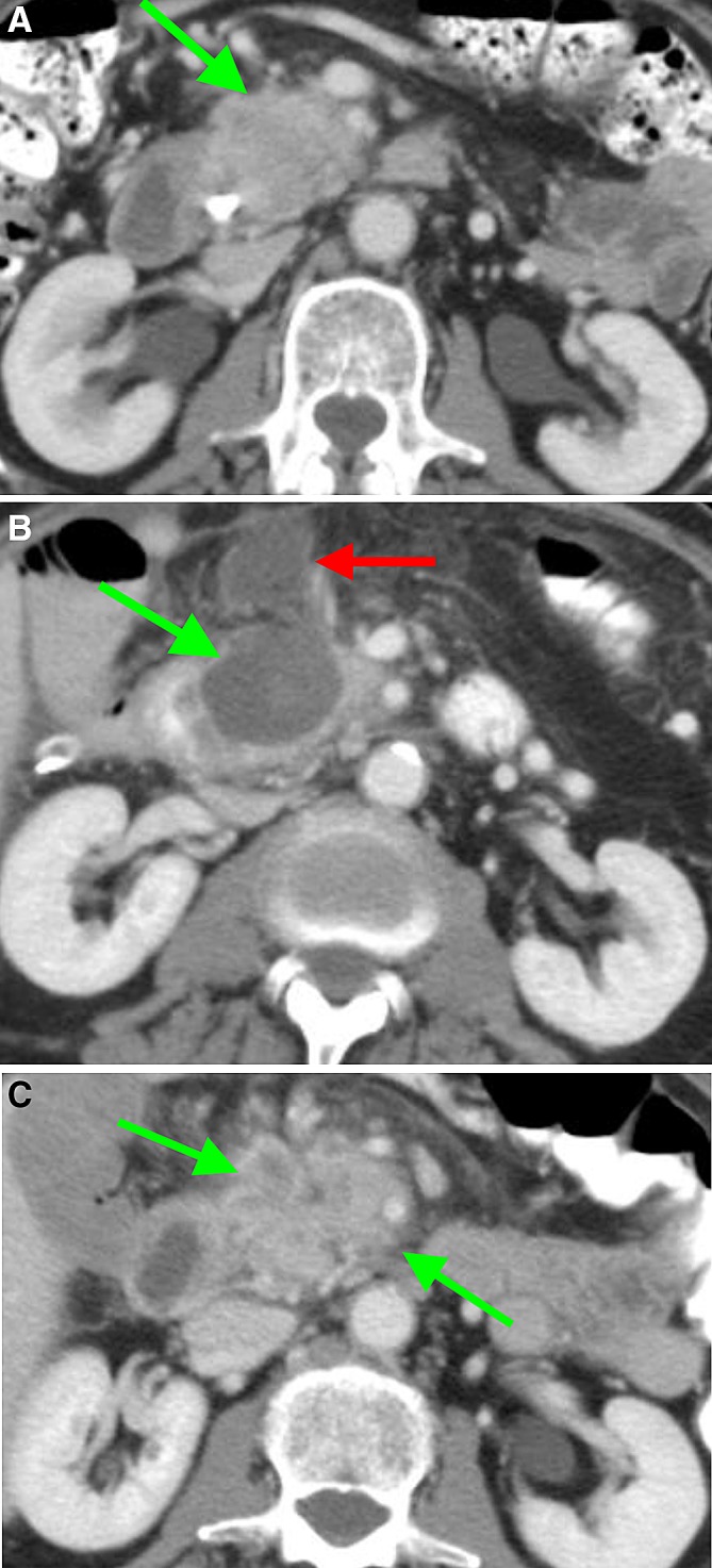
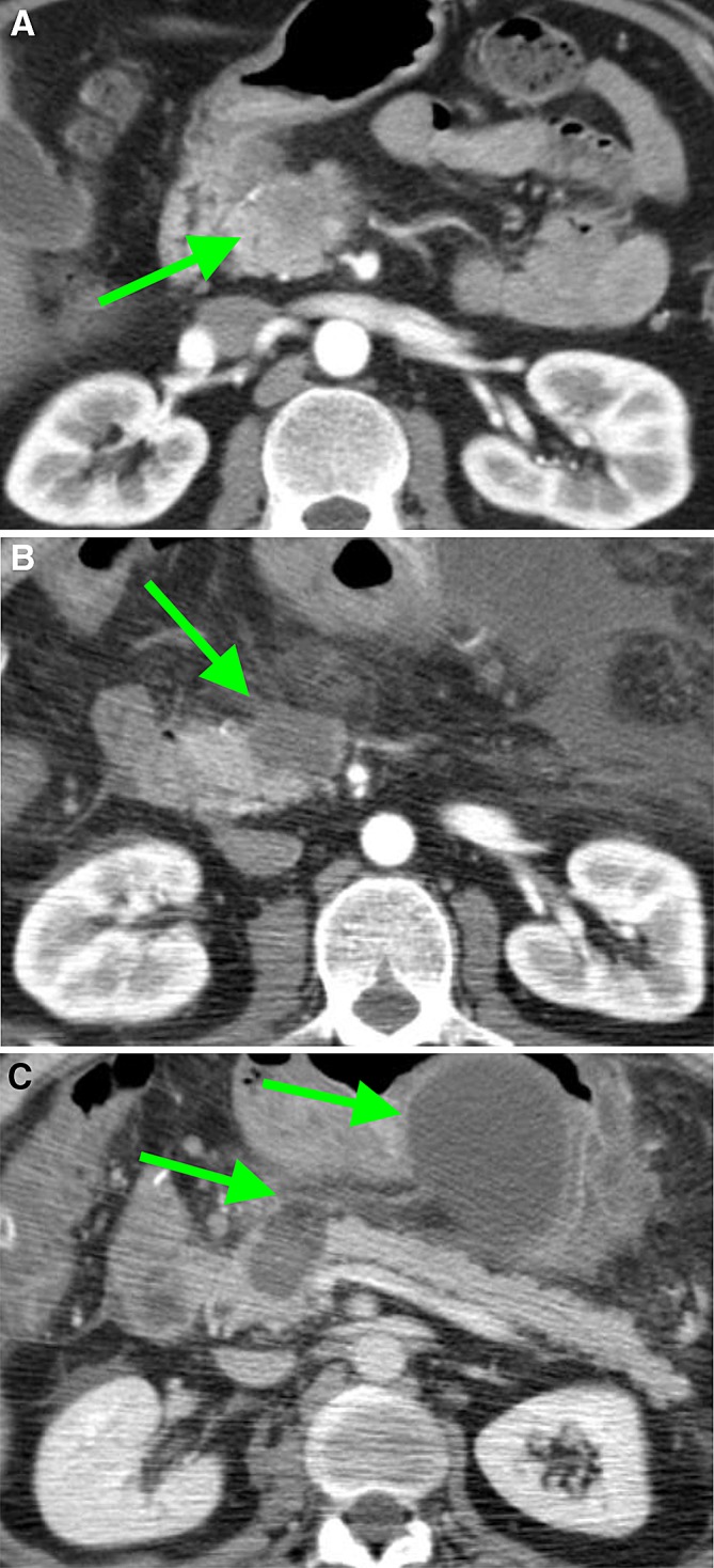
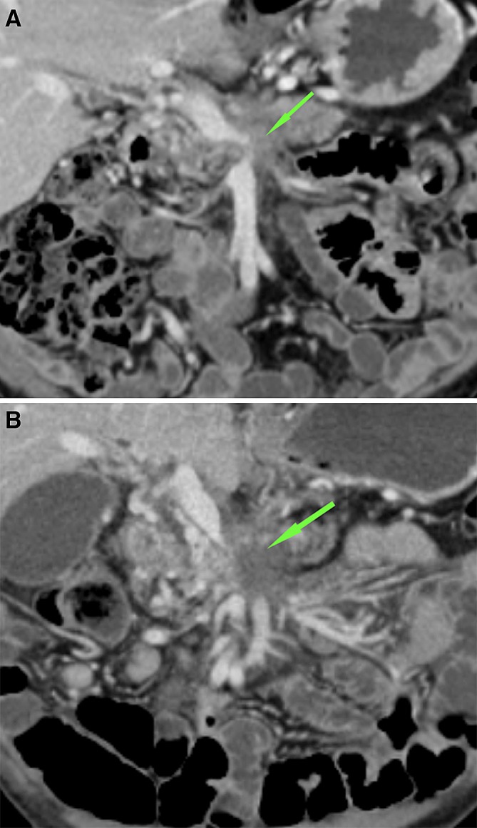
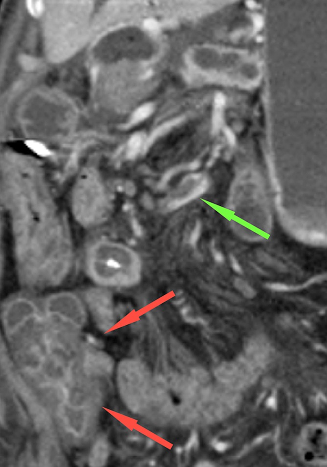
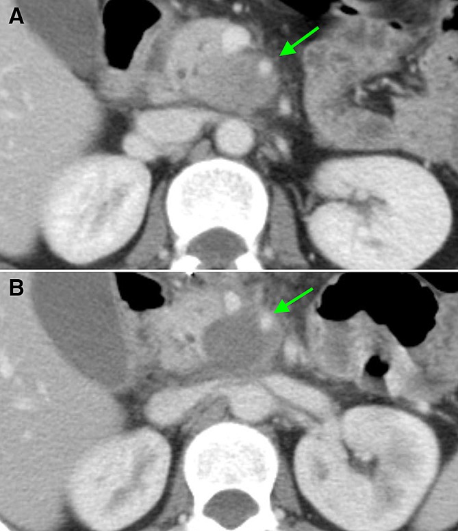
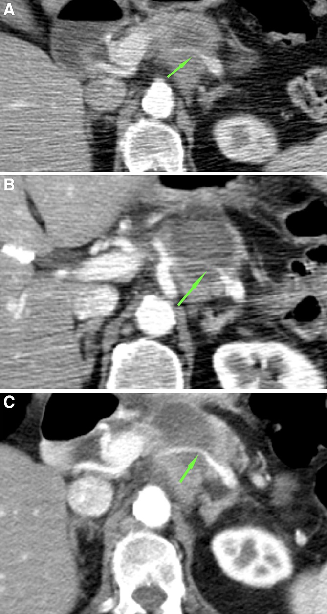
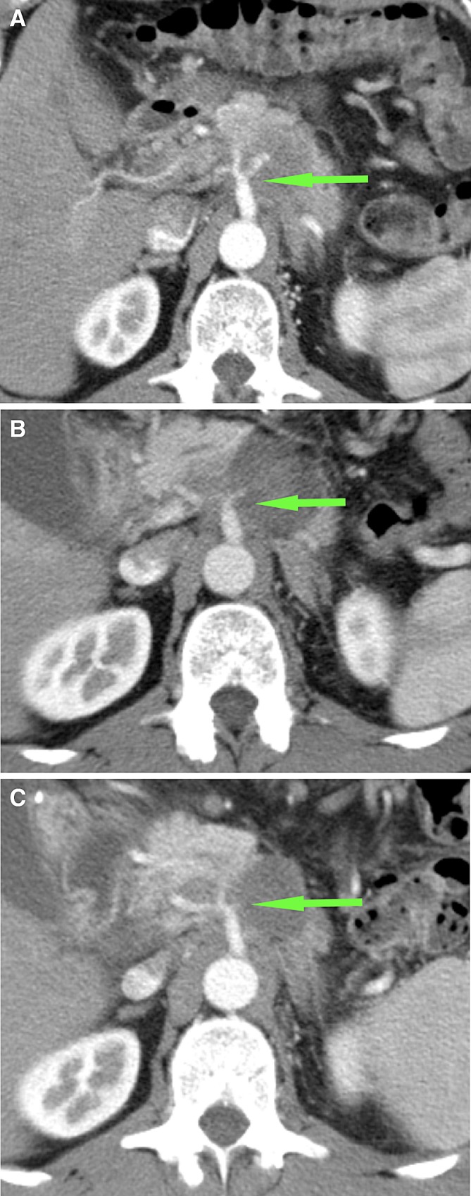
Results: 51 CT-scans were assessed. One week after RFA, the ablation zone was visible in all patients as a (partially) sharply defined (83%), heterogeneous area (94%). At 3 months of follow-up, the ablation zone was completely invaded by tumor in 67% of patients and still present, but decreased in 33%. In two patients (11%), local thrombosis and/or occlusion of the superior mesenteric vein occurred. The occlusions persisted without clinical consequences and the thrombosis disappeared. A peripancreatic fluid collection was visible 1 week after RFA in 3 patients, wherein the ablation zone extended ventrally outside of the pancreas.

Conclusions: Directly after RFA for LAPC, a well-defined ablation zone is visible on CT-imaging. This ablation zone is usually replaced by tumor ingrowth after 3 months. Moreover, the ablation zone regularly included vascular structures, with rare asymptomatic venous occlusion or thrombosis and without adverse effects on arteries.

Keywords: Computed tomography; Imaging findings; Locally advanced pancreatic cancer; Radiofrequency ablation.


Conflict of interest statement

All the authors declare that they have no conflict of interest.

Figures

Fig. 1
Fig. 1
CT-images in the axial plane, of a 74-year-old woman with A a ductal adenocarcinoma in the head of the pancreas (arrow) on the pre-RFA scan, showing 90°–180° circumferential involvement of the superior mesenteric artery. B One-week post RFA, a sharply demarcated ablation zone is visible as a heterogeneous area (arrow). Moreover, the ablation zone extends ventrally outside the pancreas contour and a fluid collection is present in the transverse mesocolon. C Tumor replacing the ablation zone (arrow) at 3 months post-RFA. Note the inhomogeneous enhancement, the mass effect and the increasing size and involvement of the vessels over time
Fig. 2
Fig. 2
CT-images in the axial plane, of a 56-year-old man with A a ductal adenocarcinoma in the head of the pancreas (arrow) of 3.4 cm transversal on the pre-RFA scan. B and C On CT 1-week post-RFA, the ablation zone included the entire tumor (arrow). C The ablation zone included the ventral contour of the pancreas (left arrow) with an accompanying fluid collection in the lesser sac (right arrow)
Fig. 3
Fig. 3
CT-images in the coronal plane, of the same patient as Fig. 6. A The superior mesenteric vein is already narrow on the pre-RFA imaging (arrow). B One week after RFA, the SMV is occluded for the part that is embedded in the ablation zone (arrow)
Fig. 4
Fig. 4
CT-images in the coronal plane, of a 66-year-old woman with a ductal adenocarcinoma in the head, neck and corpus of the pancreas. A The CT-image pre-RFA, shows a superior mesenteric vein (SMV) circumferential involvement of > 270°, with a pre-existent lumen reduction of > 50% (arrow) and B no thrombus present (arrow). C On the CT-image 1-week post-RFA the SMV and portal vein are occluded within the ablation zone and D a local thrombus is present in the SMV (arrow)
Fig. 5
Fig. 5
CT-image in the coronal plane, 1-week post-RFA, of a 68-year-old woman with a ductal adenocarcinoma located in the head and neck of the pancreas, showing a thrombus in the ileocolic vein (green arrow), resulting in edema of the ascending colon (red arrows)
Fig. 6
Fig. 6
CT-images in the axial plane, of a 44-year-old woman with a ductal adenocarcinoma in the head of the pancreas. A On the pre-RFA CT-image, the superior mesenteric artery (SMA) was involved in the tumor, with a circumferential involvement of > 270, with normal patency and without anatomical changes. B On the CT-image 1-week post-RFA, the SMA was included in the ablation zone for 90°–180°, without the occurrence of anatomical changes
Fig. 7
Fig. 7
CT-images in the axial plane, of a 76-year-old woman with a ductal adenocarcinoma in the pancreatic corpus. A The splenic artery is encased by the tumor but still patent(arrow). B One week after RFA, the splenic artery is in contact with the ablation zone, but still patent (arrow). C This remained unchanged at follow-up 1 month later (arrow)
Fig. 8
Fig. 8
CT-images in the axial plane, of a 51-year-old man with a ductal adenocarcinoma in the corpus of the pancreas. A On the CT-imaging before RFA, the coeliac trunk and hepatic artery are closely related to the tumor. B One week after the procedure, these arteries are constricted but still patent (arrow). C This remained stable during follow-up 3 months later (arrow)

References

    1. Ferlay J, Soerjomataram I, Ervik M, Eser S, Mathers C, Rebelo M, et al. Cancer incidence and mortality worldwide: sources, methods and major patterns in GLOBOCAN 2012. Int J Cancer. 2015;136:359–386. doi: 10.1002/ijc.29210. - DOI - PubMed
    1. Siegel RL, Miller KD, Jemal A. Cancer statistics. CA Cancer J Clin. 2016;66:7–30. doi: 10.3322/caac.21332. - DOI - PubMed
    1. Ducreux M, Cuhna AS, Caramella C, Hollebecque A, Burtin P, Goéré D, et al. Cancer of the pancreas: ESMO Clinical Practice Guidelines for diagnosis, treatment and follow-up. Ann Oncol. 2015;26:56–68. doi: 10.1093/annonc/mdv295. - DOI - PubMed
    1. Rombouts SJE, Vogel JA, Van Santvoort HC, van Lienden KP, van Hillegersberg R, Busch OR, et al. Systematic review of innovative ablative therapies for the treatment of locally advanced pancreatic cancer. Br J Surg. 2015;102(3):182–193. doi: 10.1002/bjs.9716. - DOI - PubMed
    1. Feldman MK, Gandhi NS. Imaging evaluation of pancreatic cancer. Surg Clin N Am. 2016;96(6):1235–1256. doi: 10.1016/j.suc.2016.07.007. - DOI - PubMed