Review
. 2019;26(16):2918-2932.
doi: 10.2174/0929867325666180228165527.

Potential Roles for G-Quadruplexes in Mitochondria

Affiliations
Review

Potential Roles for G-Quadruplexes in Mitochondria

Micol Falabella et al. Curr Med Chem. 2019.

Abstract

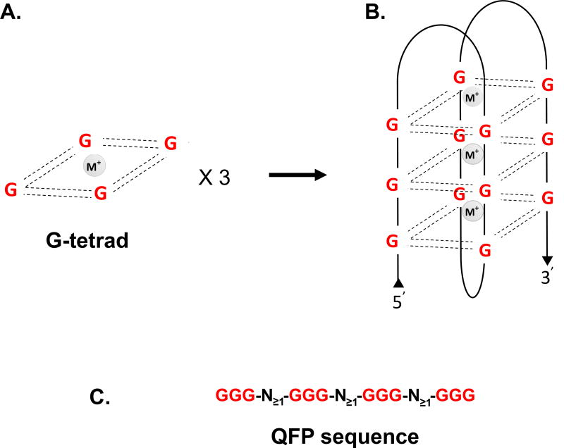
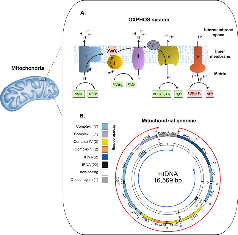
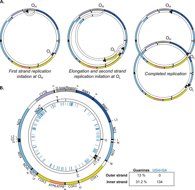
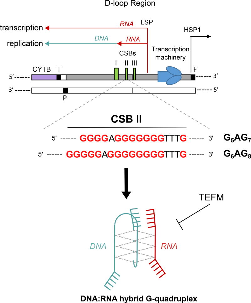
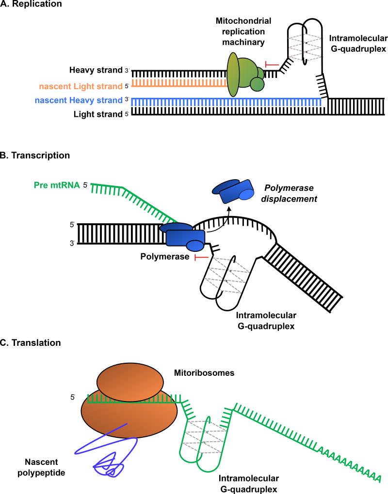
Some DNA or RNA sequences rich in guanine (G) nucleotides can adopt noncanonical conformations known as G-quadruplexes (G4). In the nuclear genome, G4 motifs have been associated with genome instability and gene expression defects, but they are increasingly recognized to be regulatory structures. Recent studies have revealed that G4 structures can form in the mitochondrial genome (mtDNA) and potential G4 forming sequences are associated with the origin of mtDNA deletions. However, little is known about the regulatory role of G4 structures in mitochondria. In this short review, we will explore the potential for G4 structures to regulate mitochondrial function, based on evidence from the nucleus.

Keywords: G-quadruplexes; G4 ligand; mitochondrial gene expression; mitochondrial genome instability; mtDNA; mtDNA deletions.; mtDNA depletion.


Figures

Figure 1
Figure 1. G-quadruplex structure
(A) Four guanines (G) form planar cyclic conformations through eight Hoogsteen-hydrogen bonds (dashed lines) and stabilized by the presence of a central metal cation (M+). (B) G4 structure in antiparallel conformation. Numerous topologies are possible. (C) From the requirement of four runs of guanine, QFP sequences can be predicted within the same strand. Traditional prediction requires at least two G per run and one nucleotide to form a loop. Shown is a prediction scheme typically used for identifying three guanine stack G4-forming potential (QFP) sequences. Limits to size of window for search usually range from 30–50 bp.
Figure 2
Figure 2. Schematic representation of the oxidative phosphorylation (OXPHOS) system and mitochondrial DNA (mtDNA)
(A) Main components of the mitochondrial OXPHOS system: Complex I (blue), Coenzyme Q (CoQ; pink), Complex II (orange), Complex III (purple), Cytochrome C (CytC; grey), Complex IV (yellow) and Complex V (red). The redox metabolites are shown in green boxes and the final OXPHOS reaction is represented in red boxes. (B) The mitochondrial genome is a 16,569 bp circular double-stranded DNA molecule that contains 37 genes encoding for 13 OXPHOS proteins (colors matched panel A) and the tRNAs (black) and rRNAs (dark blue) required for their expression. The number of genes per group are indicated in parenthesis. The genes are encoded on either strand, shown with the two strands separated to indicate the relevant coding sequences on each polycistronic pre-mRNA transcript (indicated by red and blue arrows).
Figure 3
Figure 3. Mitochondrial genome replication and predicted G4 forming potential sequences
(A) Simplified diagram of mtDNA asynchronous replication. Left: first strand replication initiation at ori-H (OH). DNA starts as duplex, with synthesis of the heavy strand initiated in the heavy strand origin (OH). Middle: elongation of the first strand and initiation of the second strand replication at ori-L (OL). Continuous elongation of the first strand displaces the parental heavy strand of mtDNA with nascent DNA. Once replication crosses the primary second strand origin (OL), replication in the other direction begins. Right: Elongation of both strands continues until two semi-conservative molecules are generated. Note that only the heavy strand is single stranded in this process. (B) Representation of the G4-forming sequences in the heavy strand (inner strand; 31.2% guanines) of the human mtDNA. The figure represents validated unstable G4 (UG4; n=63) and G4 (n=71) out of 209 sequences experimentally tested.
Figure 4
Figure 4. Mitochondrial control region and transcription termination induced by G4 structure
The light strand promoter (LSP; red) transcription can be arrested by a hybrid DNA:RNA G4 structure formed at the conserved block sequence II (CSB II). The arrested RNA is thought to serve as a primer to initiate the leading-strand DNA (green) replication. Two alternative regions at the CSB II could lead to the G4 formation, the G5AG7 (rare polymorphism) and the G6AG8 (most genomes) sequences. The mitochondrial transcription elongation factor (TFEM) prevents the CSB II transcription termination.
Figure 5
Figure 5. Potential G4 formation during mtDNA replication, transcription and translation
G4 structures can form in single-stranded nucleic acids. (A) During mtDNA replication, the displaced G-rich heavy strand is a potential hotspot for the G4 formation, which would arrest the mitochondrial replication machinery (green) during second strand synthesis. (B) A model of transcription inhibition by G4 structures. The unresolved G4 structures would occur in the mtDNA template strand preventing the polymerase (blue) activity. (C) A model of translation inhibition by G4 structures forming in the mRNA.

References

    1. Burge S, Parkinson GN, Hazel P, Todd AK, Neidle S. Quadruplex DNA: sequence, topology and structure. Nucleic Acids Res. 2006;34:5402–15. - PMC - PubMed
    1. Sen D, Gilbert W. Formation of parallel four-stranded complexes by guanine-rich motifs in DNA and its implications for meiosis. Nature. 1988;334:364–366. - PubMed
    1. Sen D, Gilbert W. A sodium-potassium switch in the formation of four-stranded G4-DNA. Nature 1990 Mar 29; - PubMed
    1. Hazel P, Parkinson GN, Neidle S. Topology variation and loop structural homology in crystal and simulated structures of a bimolecular DNA quadruplex. J. Am. Chem. Soc. 2006;128:5480–7. - PubMed
    1. Karsisiotis AI, Hessari NM, Novellino E, Spada GP, Randazzo A, Webba da Silva M. Topological characterization of nucleic acid G-quadruplexes by UV absorption and circular dichroism. Angew. Chem. Int. Ed. Engl. 2011;50:10645–8. - PubMed

LinkOut - more resources