Review
. 2018 Jun 1;314(6):G668-G676.
doi: 10.1152/ajpgi.00026.2018. Epub 2018 Mar 1.

Biliverdin reductase and bilirubin in hepatic disease

Affiliations
Review

Biliverdin reductase and bilirubin in hepatic disease

Lauren Weaver et al. Am J Physiol Gastrointest Liver Physiol. .

Abstract

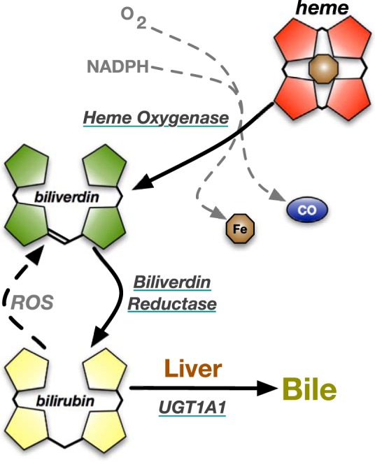
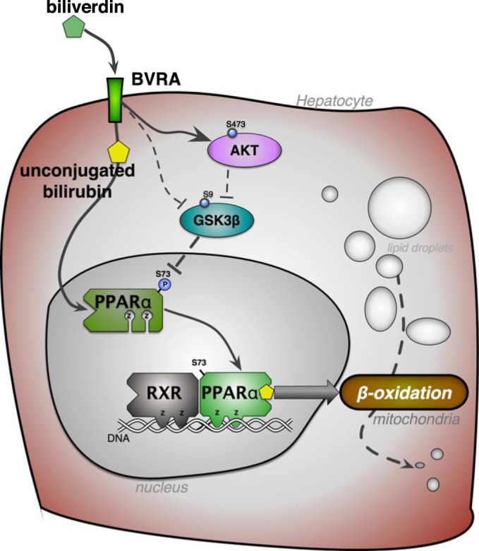
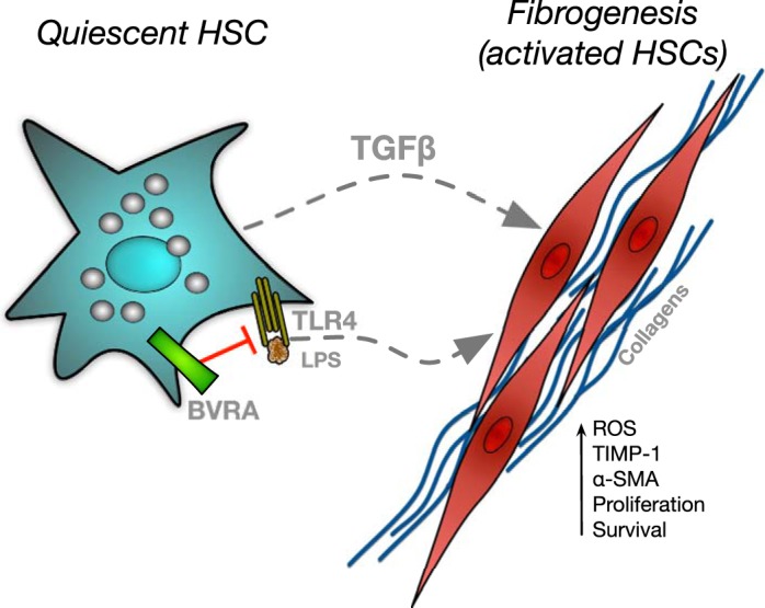
The buildup of fat in the liver (hepatic steatosis) is the first step in a series of incidents that may drive hepatic disease. Obesity is the leading cause of nonalcoholic fatty liver disease (NAFLD), in which hepatic steatosis progresses to liver disease. Chronic alcohol exposure also induces fat accumulation in the liver and shares numerous similarities to obesity-induced NAFLD. Regardless of whether hepatic steatosis is due to obesity or long-term alcohol use, it still may lead to hepatic fibrosis, cirrhosis, or possibly hepatocellular carcinoma. The antioxidant bilirubin and the enzyme that generates it, biliverdin reductase A (BVRA), are components of the heme catabolic pathway that have been shown to reduce hepatic steatosis. This review discusses the roles for bilirubin and BVRA in the prevention of steatosis, their functions in the later stages of liver disease, and their potential therapeutic application.

Keywords: ALD; BVRA; NAFLD; PPARα; bilirubin; fatty liver disease; fibrosis.


Figures

Fig. 1.
Fig. 1.
Heme catabolic pathway. Heme contains a porphyrin ring and a central iron atom. In heme degradation, heme oxygenase breaks open the porphyrin ring with NADPH and oxygen (O2) resulting in the release of iron (Fe), carbon monoxide (CO), and biliverdin. Biliverdin is reduced by biliverdin reductase to bilirubin. Reactive oxygen species (ROS) can recycle bilirubin back to biliverdin. In the liver, bilirubin is conjugated with glucuronic acid by UDP-glucuronosyltransferase 1A1 (UGT1A1) and excreted into the bile.
Fig. 2.
Fig. 2.
Biliverdin reductase signaling reduces hepatic fat accumulation. Biliverdin reductase A (BVRA) generation of unconjugated bilirubin from biliverdin activates peroxisome proliferator-activated receptor-α (PPARα; 99), which is a transcription factor that interacts with the retinoid X receptor (RXR) to increase genes in the β-oxidation pathway that burn fatty acids in the mitochondria. BVRA activates Akt, which inhibits glycogen synthase kinase-3β (GSK3β) through increased phosphorylation at serine 9 (S9) to prevent hepatic steatosis (37). BVRA can also directly interact with GSK3β to regulate activity (69). Inhibition of GSK3β results in increased activity of PPARα via decreased phosphorylation (P) at serine 73 (37). Z, zinc finger DNA binding domains. The signaling functions of BVRA and bilirubin have been reviewed further by Hamoud et al. (34), O’Brien et al. (76), and Rochette et al. (86).
Fig. 3.
Fig. 3.
Proposed inhibitory role of biliverdin reductase A (BVRA) in fibrogenesis. Hepatic stellate cells (HSCs) are activated by lipopolysaccharide (LPS) binding to toll-like receptor 4 (TLR4) or stimulation by transforming growth factor-β (TGF-β). Activated HSCs increase collagen production as well as several other factors to promote liver fibrogenesis. BVRA can attenuate fibrogenesis through antagonism of TLR4. α-SMA, α-smooth muscle actin; ROS, reactive oxygen species; TIMP-1, tissue inhibitor of metalloproteinases 1.

References

    1. Abbasi A, Deetman PE, Corpeleijn E, Gansevoort RT, Gans RO, Hillege HL, van der Harst P, Stolk RP, Navis G, Alizadeh BZ, Bakker SJ. Bilirubin as a potential causal factor in type 2 diabetes risk: a Mendelian randomization study. Diabetes 64: 1459–1469, 2015. doi:10.2337/db14-0228. - DOI - PMC - PubMed
    1. Ahmad Z, Salim M, Maines MD. Human biliverdin reductase is a leucine zipper-like DNA-binding protein and functions in transcriptional activation of heme oxygenase-1 by oxidative stress. J Biol Chem 277: 9226–9232, 2002. doi:10.1074/jbc.M108239200. - DOI - PubMed
    1. Baranano DE, Rao M, Ferris CD, Snyder SH. Biliverdin reductase: a major physiologic cytoprotectant. Proc Natl Acad Sci USA 99: 16093–16098, 2002. doi:10.1073/pnas.252626999. - DOI - PMC - PubMed
    1. Bataller R, Brenner DA. Liver fibrosis. J Clin Invest 115: 209–218, 2005. doi:10.1172/JCI24282. - DOI - PMC - PubMed
    1. Belo L, Nascimento H, Kohlova M, Bronze-da-Rocha E, Fernandes J, Costa E, Catarino C, Aires L, Mansilha HF, Rocha-Pereira P, Quintanilha A, Rêgo C, Santos-Silva A. Body fat percentage is a major determinant of total bilirubin independently of UGT1A1*28 polymorphism in young obese. PLoS One 9: e98467, 2014. doi:10.1371/journal.pone.0098467. - DOI - PMC - PubMed

Publication types

MeSH terms