Review
. 2018 Jun 20:87:239-261.
doi: 10.1146/annurev-biochem-062917-012405. Epub 2018 Mar 1.

Translesion and Repair DNA Polymerases: Diverse Structure and Mechanism

Affiliations
Review

Translesion and Repair DNA Polymerases: Diverse Structure and Mechanism

Wei Yang et al. Annu Rev Biochem. .

Abstract

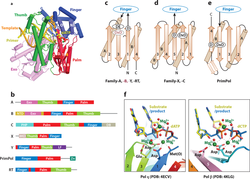
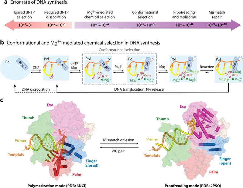
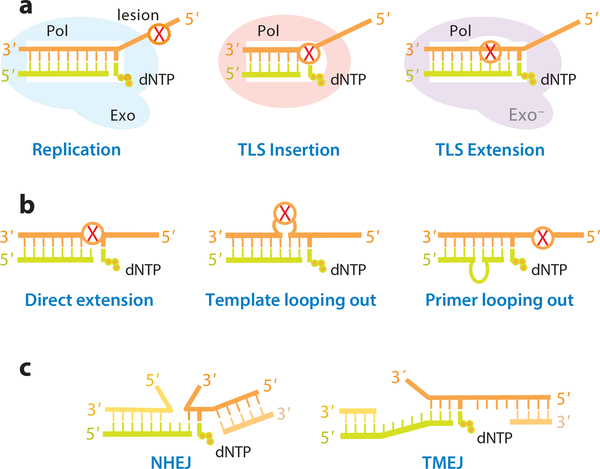
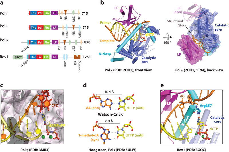
The number of DNA polymerases identified in each organism has mushroomed in the past two decades. Most newly found DNA polymerases specialize in translesion synthesis and DNA repair instead of replication. Although intrinsic error rates are higher for translesion and repair polymerases than for replicative polymerases, the specialized polymerases increase genome stability and reduce tumorigenesis. Reflecting the numerous types of DNA lesions and variations of broken DNA ends, translesion and repair polymerases differ in structure, mechanism, and function. Here, we review the unique and general features of polymerases specialized in lesion bypass, as well as in gap-filling and end-joining synthesis.

Keywords: A-family; B-family; DSBs; NHEJ; TLS; TMEJ; X-family; Y-family; proofreading; sGRS; small gap-filling repair synthesis.


Figures

Figure 1
Figure 1
Architecture of DNA polymerases. (a) A DNA polymerase is composed of thumb (green), finger (blue) and palm domain (red). Replicative polymerases often include an intrinsic proofreading 3´−5´ exonuclease (light purple). The ternary complex of an A-family DNA polymerase (PDB: 1LV5) is shown as an example. (b) Primary structures of seven DNA polymerase families. (c-e) Topology diagrams of the palm domain in A, B, Y, and RT families, C and X families, and PrimPol. (f) Side-by-side view of the catalytic centers of Pol η and Pol β.
Figure 2
Figure 2
Basis for DNA polymerase fidelity. (a) Determinants of the error rates of DNA polymerases. (b) Conformational and Mg2+-mediated chemical selection in the DNA synthesis reaction. The requirement for three Mg2+ ions is universal, and conformational selection (boxed) occurs in replicative and a few repair polymerases. Mg2+A binding and conformational change can occur without one another in replicative polymerases. (c) In the DNA synthesis (finger-closed) mode, a replicative polymerase (B-family RB69 as an example, PDB: 3NCI) interacts with the DNA and nascent base pair seamlessly. In the proofreading mode (PDB: 2P5O), DNA is detached from the open finger and disordered thumb, and primer strand is partially separated from the template and migrates to the Exo active site for cleavage.
Figure 3
Figure 3
Mechanisms for DNA translesion and repair synthesis. (a) Diagram of insertion and extension during TLS. (b) Three possible ways to achieve TLS extension. (c) Discontinuous strands of broken DNA can be accommodated similarly to looping out.
Figure 4
Figure 4
Mechanism of TLS insertion by Y-family polymerases. (a) Primary structures of human Y-family polymerases. (b) Crystal structures of Pol κ. In the ternary complex (PDB: 2OH2), the N-clasp stabilizes the LF and the structural gap between the catalytic core and LF to accommodate a bulky adduct. Without DNA the LF is flexible (PDB: 1T94). (c) Crystal structures of Pol η accommodating a thymine dimer in the active site (PDB: 3MR3). (d) The Pol ι active site favors a narrow Hoogsteen basepair (PDB: 5ULW) and not WC pair. (e) Crystal structure of Rev1, which utilizes an Arg sidechain to select dCTP for incorporation (PDB: 3GQC). The N-digit is inserted between the LF and catalytic core.
Figure 5
Figure 5
TLS extension by Pol ζ and E. coli Pol II. (a) Primary structures of E. coli Pol II, yeast and human Rev3. (b) Two nucleotides including an AP site (THF) on the template strand are looped out in complex with E. coli Pol II (PDB: 3K5M).
Figure 6
Figure 6
TLS and end joining by Pol ν and θ. (a) Primary structures of human Pol ν and θ. (b) Ins 2 in Pol ν results in the cavity and a flexible thumb for primer looping out (PDB:4XVK, 4XVM).
Figure 7
Figure 7
sGRS by the X-family polymerases. (a) Primary structures of Pol β, λ, μ and TdT. (b) Crystal structure of the Pol β-DNA-dNTP complex (PDB: 2FMS). Domains are labeled according to the right-hand convention. In the left-hand definition, finger and thumb would be swapped. (c) Gap filling synthesis and dRP removal by Pol β. The two DNA segments are at ~90° angle. (d) Pol λ can bridge two DNA ends by one base pair and incorporate a dNTP to match the trans template. (e) Structure of TdT bridging two DNA ends without base pairing (PDB: 4QZB). (f) Pol μ can bridge two DNA ends by a mismatched base pair and incorporate a dNTP that matches the trans template. Loop1 insertion in TdT and Pol μ segregates the two base pairs in the active site from upstream DNA.

References

    1. Bessman MJ, Kornberg A, Lehman IR, Simms ES. 1956. Enzymic synthesis of deoxyribonucleic acid. Biochim Biophys Acta 21: 197–8 - PubMed
    1. Englund PT, Deutscher MP, Jovin TM, Kelly RB, Cozzarelli NR, Kornberg A. 1968. Structural and functional properties of Escherichia coli DNA polymerase. Cold Spring Harb Symp Quant Biol 33: 1–9 - PubMed
    1. Ollis DL, Brick P, Hamlin R, Xuong NG, Steitz TA. 1985. Structure of large fragment of Escherichia coli DNA polymerase I complexed with dTMP. Nature 313: 762–6 - PubMed
    1. Lindahl T 2001. Keynote: past, present, and future aspects of base excision repair. Prog Nucleic Acid Res Mol Biol 68: xvii–xxx - PubMed
    1. Setlow RB. 1966. Cyclobutane-type pyrimidine dimers in polynucleotides. Science 153: 379–86 - PubMed

Publication types

MeSH terms

Substances