. 2018 Mar 1;23(3):549.
doi: 10.3390/molecules23030549.

Trimethylamine-N-Oxide (TMAO)-Induced Impairment of Cardiomyocyte Function and the Protective Role of Urolithin B-Glucuronide

Affiliations

Trimethylamine-N-Oxide (TMAO)-Induced Impairment of Cardiomyocyte Function and the Protective Role of Urolithin B-Glucuronide

Monia Savi et al. Molecules. .

Abstract

One of the most recently proposed candidates as a potential trigger for cardiovascular diseases is trimethylamine-N-oxide (TMAO). Possible direct effects of TMAO on myocardial tissue, independent of vascular damage, have been only partially explored so far. In the present study, we assessed the detrimental direct effects of TMAO on cardiomyocyte contractility and intracellular calcium dynamics, and the ability of urolithin B-glucuronide (Uro B-gluc) in counteracting TMAO-induced cell damage. Cell mechanics and calcium transients were measured, and ultrastructural analysis was performed in ventricular cardiomyocytes isolated from the heart of normal adult rats. Cells were either untreated, exposed to TMAO, or to TMAO and Uro B-gluc. TMAO exposure worsened cardiomyocyte mechanics and intracellular calcium handling, as documented by the decrease in the fraction of shortening (FS) and the maximal rate of shortening and re-lengthening, associated with reduced efficiency in the intracellular calcium removal. Ultrastructurally, TMAO-treated cardiomyocytes also exhibited glycogen accumulation, a higher number of mitochondria and lipofuscin-like pigment deposition, suggesting an altered cellular energetic metabolism and a higher rate of protein oxidative damage, respectively. Uro B-gluc led to a complete recovery of cellular contractility and calcium dynamics, and morphologically to a reduced glycogen accumulation. We demonstrated for the first time a direct negative role of TMAO on cardiomyocyte functional properties and the ability of Uro B-gluc in counteracting these detrimental effects.

Keywords: Trimethylamine-N-oxide; cardiomyocyte mechanics; cardiovascular diseases; ellagitannins; urolithins.


Conflict of interest statement

The authors declare no conflict of interest.

Figures

Figure 1
Figure 1
Cell mechanics. Representative examples of sarcomere shortening (SL) (A) recorded from CTRL, TMAO100, TMAO20 and TMAO20 + Uro B-gluc ventricular myocytes. In bar graphs (BF): mean values ± SEM of diastolic sarcomere length (SL; B), maximal rate of shortening (−dl/dtmax; C), fraction of shortening (FS; D), maximal rate of re-lengthening (+dl/dtmax; E), time at 10%, 50%, and 90% of re-lengthening (TRL10%, TRL50%, TRL90%; F), measured in CTRL (n = 115), TMAO100 (n = 84), TMAO20 (n = 100), and TMAO20 + Uro B-gluc (n = 82) cells. * p < 0.05 significant differences vs. CTRL; # p < 0.05 significant differences vs. TMAO20. General Linear Model/ANOVA GLM/ANOVA for repeated measurements was used first to compare CTRL, TMAO100, and TMAO20 groups to evaluate TMAO dose-dependent effects, and then CTRL, TMAO20, and TMAO20 + Uro B-gluc groups, after selecting the TMAO minimal effective dose.
Figure 1
Figure 1
Cell mechanics. Representative examples of sarcomere shortening (SL) (A) recorded from CTRL, TMAO100, TMAO20 and TMAO20 + Uro B-gluc ventricular myocytes. In bar graphs (BF): mean values ± SEM of diastolic sarcomere length (SL; B), maximal rate of shortening (−dl/dtmax; C), fraction of shortening (FS; D), maximal rate of re-lengthening (+dl/dtmax; E), time at 10%, 50%, and 90% of re-lengthening (TRL10%, TRL50%, TRL90%; F), measured in CTRL (n = 115), TMAO100 (n = 84), TMAO20 (n = 100), and TMAO20 + Uro B-gluc (n = 82) cells. * p < 0.05 significant differences vs. CTRL; # p < 0.05 significant differences vs. TMAO20. General Linear Model/ANOVA GLM/ANOVA for repeated measurements was used first to compare CTRL, TMAO100, and TMAO20 groups to evaluate TMAO dose-dependent effects, and then CTRL, TMAO20, and TMAO20 + Uro B-gluc groups, after selecting the TMAO minimal effective dose.
Figure 2
Figure 2
Intracellular calcium transients. Representative examples of calcium transients (A), normalized traces: fold increase, f/f0) recorded from CTRL, TMAO100, TMAO20 and TMAO20 + Uro B-gluc ventricular myocytes. In bar graphs (BD): mean values ± SEM of Ca2+ transient amplitude expressed as peak fluorescence normalized to baseline fluorescence (f/f0; B), time to peak of the calcium transient (TTP; C), and time constant of the intracellular calcium decay (tau; D), measured in CTRL (n = 50), TMAO100 (n = 33), TMAO20 (n = 35), and TMAO20 + Uro B-gluc (n = 29) cardiomyocytes. * p < 0.05 significant differences vs. CTRL; # p < 0.05 significant differences vs. TMAO20 (GLM/ANOVA, other explanations as in Figure 1).
Figure 3
Figure 3
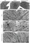
Effects of TMAO and TMAO + Uro B-gluc on cardiomyocyte ultrastructure (TEM study). (AC): low magnification TEM images of isolated cardiomyocytes in the absence (A) or presence of TMAO alone (B) or in combination with Uro B-gluc (C). (D,E): CTRL untreated cardiomyocytes showing a regular distribution of mitochondria and glycogen (arrows) between sarcomeric units; (F,G): the in vitro exposure of cardiomyocytes to TMAO induced accumulation of glycogen (arrows), occasional lipofuscin-like deposition (LD) and mitochondria enlargement. (H,I): the addition of Uro B-gluc to TMAO reduced glycogen compartmentalization without changes in mitochondrial size compared to TMAO alone. M: Mitochondria; N: cardiomyocytenucleus. Scale bars: 2 µm (AD,F,H); 1 µm (E,G,I).
Figure 4
Figure 4
Effects of TMAO and TMAO + Uro B-gluc on cardiomyocyte glycogen content and mitochondria. Mean values ± SEM of the volume fraction of glycogen (A, %) and the number of mitochondria (B, n/10 μm2) in CTRL, TMAO20, and TMAO20 + Uro B-gluc cells. * p < 0.05 significant differences vs. CTRL; # p < 0.05 significant differences vs. TMAO20 (non-parametric statistical test: Kruskal Wallis and U-Mann Whitney test).

References

    1. World Health Organization (WHO) Global Status Report on Noncommunicable Diseases 2014. WHO; Geneva, Switzerland: 2014.
    1. Stock J. Gut microbiota: An environmental risk factor for cardiovascular disease. Atherosclerosis. 2013;229:440–442. doi: 10.1016/j.atherosclerosis.2013.05.019. - DOI - PubMed
    1. Ufnal M., Zadlo A., Ostaszewski R. TMAO: A small molecule of great expectations. Nutrition. 2015;31:1317–1323. doi: 10.1016/j.nut.2015.05.006. - DOI - PubMed
    1. Li X.S., Obeid S., Klingenberg R., Gencer B., Mach F., Raber L., Windecker S., Rodondi N., Nanchen D., Muller O., et al. Gut microbiota-dependent trimethylamine N-oxide in acute coronary syndromes: A prognostic marker for incident cardiovascular events beyond traditional risk factors. Eur. Heart J. 2017;38:814–824. doi: 10.1093/eurheartj/ehw582. - DOI - PMC - PubMed
    1. Meyer K.A., Benton T.Z., Bennett B.J., Jacobs D.R., Jr., Lloyd-Jones D.M., Gross M.D., Carr J.J., Gordon-Larsen P., Zeisel S.H. Microbiota-Dependent Metabolite Trimethylamine N-Oxide and Coronary Artery Calcium in the Coronary Artery Risk Development in Young Adults Study (CARDIA) J. Am. Heart Assoc. 2016;5:e003970. doi: 10.1161/JAHA.116.003970. - DOI - PMC - PubMed