

A spontaneous model of spondyloarthropathies that develops bone loss and pathological bone formation: A process regulated by IL27RA-/- and mutant-p53
- PMID: 29494633
- PMCID: PMC5832250
- DOI: 10.1371/journal.pone.0193485
A spontaneous model of spondyloarthropathies that develops bone loss and pathological bone formation: A process regulated by IL27RA-/- and mutant-p53
Abstract
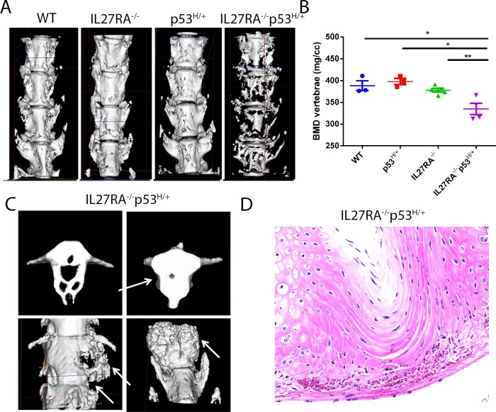
Spondyloarthropathies, the second most frequently occurring form of chronic inflammatory arthritis, affects young adults in particular. However, a proper model with which to study the biology of this disease and to develop therapeutics is lacking. One of the most accepted animal models for this disease uses HLA-B27/Hu-β2m transgenic rats; however, only 30%-50% of male HLA-B27/Hu-β2m rats develop spontaneous, clinically apparent spondylitis and have a variable time until disease onset. Here, we report a high-incidence, low-variation spontaneous mouse model that delineates how the combination of inflammatory cytokine interleukin-27 (IL-27) signaling deficiency and mitogenic signaling (mutant p53R172H) in vivo, leads to bone loss in the vertebral bodies and ossification of the cartilage in the intervertebral discs. In this human disease-like mouse model, bone loss and pathogenic bone development are seen as early as 4 months of age in the absence of inflammatory aggregates in the enthesis or intervertebral disc.
Conflict of interest statement
Figures


References
-
- Tran TM, Dorris ML, Satumtira N, Richardson JA, Hammer RE, Shang J, et al. Additional human beta2-microglobulin curbs HLA-B27 misfolding and promotes arthritis and spondylitis without colitis in male HLA-B27-transgenic rats. Arthritis Rheum. 2006;54(4):1317–27. doi: 10.1002/art.21740 . - DOI - PubMed
-
- Sherlock JP, Joyce-Shaikh B, Turner SP, Chao CC, Sathe M, Grein J, et al. IL-23 induces spondyloarthropathy by acting on ROR-gamma t(+) CD3(+)CD4(-)CD8(-) entheseal resident T cells. Nature Medicine. 2012;18(7):1069–+. doi: 10.1038/nm.2817 PubMed PMID: WOS:000306121600036. - DOI - PubMed
-
- Adamopoulos IE, Pflanz S. The emerging role of Interleukin 27 in inflammatory arthritis and bone destruction. Cytokine Growth Factor Rev. 2013;24(2):115–21. doi: 10.1016/j.cytogfr.2012.10.001 ; PubMed Central PMCID: PMCPMC3605270. - DOI - PMC - PubMed
-
- Moon SJ, Park JS, Heo YJ, Kang CM, Kim EK, Lim MA, et al. In vivo action of IL-27: reciprocal regulation of Th17 and Treg cells in collagen-induced arthritis. Exp Mol Med. 2013;45:e46 doi: 10.1038/emm.2013.89 ; PubMed Central PMCID: PMCPMC3809362. - DOI - PMC - PubMed
-
- Goldberg R, Wildbaum G, Zohar Y, Maor G, Karin N. Suppression of ongoing adjuvant-induced arthritis by neutralizing the function of the p28 subunit of IL-27. J Immunol. 2004;173(2):1171–8. . - PubMed
Publication types
MeSH terms
Substances
Grants and funding
LinkOut - more resources
Full Text Sources
Other Literature Sources
Molecular Biology Databases
Research Materials
Miscellaneous
