. 2018 Feb 24;10(2):95.
doi: 10.3390/v10020095.

A Fluorescent Cell-Based System for Imaging Zika Virus Infection in Real-Time

Affiliations

A Fluorescent Cell-Based System for Imaging Zika Virus Infection in Real-Time

Michael J McFadden et al. Viruses. .

Abstract

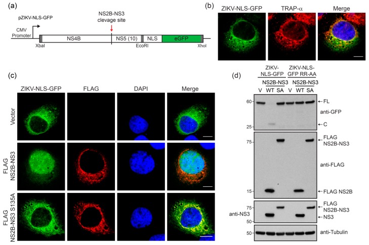
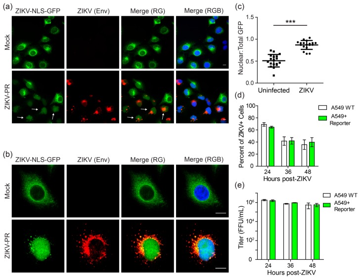
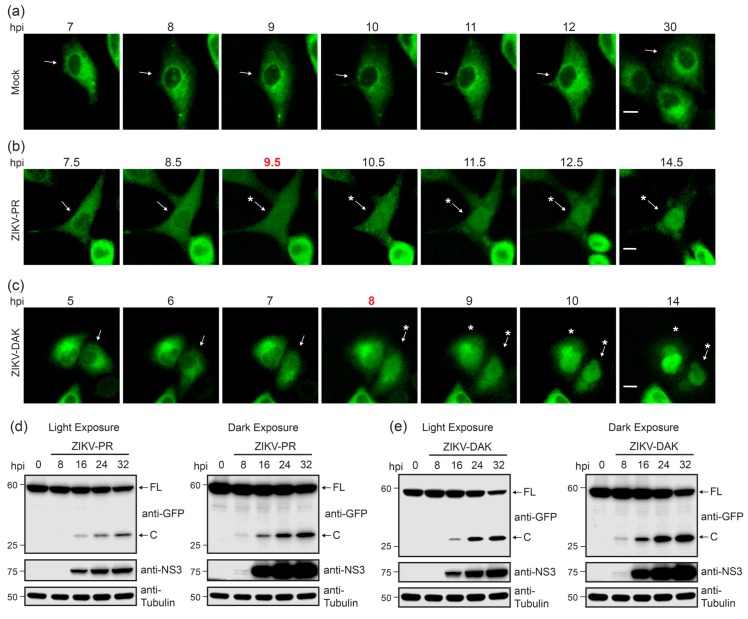
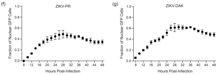
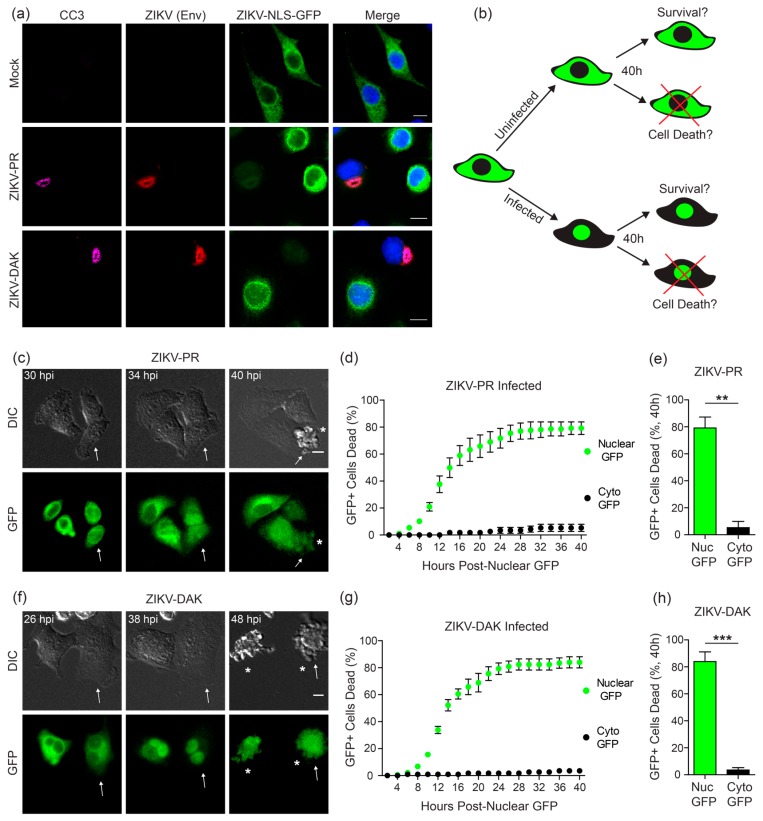
Zika virus (ZIKV) is a re-emerging flavivirus that is transmitted to humans through the bite of an infected mosquito or through sexual contact with an infected partner. ZIKV infection during pregnancy has been associated with numerous fetal abnormalities, including prenatal lethality and microcephaly. However, until recent outbreaks in the Americas, ZIKV has been relatively understudied, and therefore the biology and pathogenesis of ZIKV infection remain incompletely understood. Better methods to study ZIKV infection in live cells could enhance our understanding of the biology of ZIKV and the mechanisms by which ZIKV contributes to fetal abnormalities. To this end, we developed a fluorescent cell-based reporter system allowing for live imaging of ZIKV-infected cells. This system utilizes the protease activity of the ZIKV non-structural proteins 2B and 3 (NS2B-NS3) to specifically mark virus-infected cells. Here, we demonstrate the utility of this fluorescent reporter for identifying cells infected by ZIKV strains of two lineages. Further, we use this system to determine that apoptosis is induced in cells directly infected with ZIKV in a cell-autonomous manner. Ultimately, approaches that can directly track ZIKV-infected cells at the single cell-level have the potential to yield new insights into the host-pathogen interactions that regulate ZIKV infection and pathogenesis.

Keywords: NS2B-NS3; NS4B-NS5; Zika virus (ZIKV); apoptosis; fluorescence; live cell imaging; reporter.


Conflict of interest statement

The authors declare no conflict of interest. The founding sponsors had no role in the design of the study; in the collection, analyses, or interpretation of data; in the writing of the manuscript, and in the decision to publish the results.

Figures

Figure 1
Figure 1
A cleavable reporter to measure Zika virus (ZIKV) non-structural proteins 2B and 3 (NS2B-NS3) protease cleavage. (a) Schematic of the fluorescent ZIKV-nuclear localization signal (NLS)-GFP reporter plasmid (pZIKV-NLS-GFP) construct encoding ZIKV non-structural protein 4B (NS4B) (aa2270–2520) and the first 10 amino acids of non-structural protein 5 (NS5) (aa2521–2530), fused in frame to a nuclear localization signal (NLS) and enhanced green fluorescent protein (eGFP). The red arrow indicates the NS2B-NS3 protease cleavage site. Restriction sites used for cloning are indicated by gray boxes. (b) Confocal micrographs of Huh7 cells expressing ZIKV-NLS-GFP (green) and immunostained with the endoplasmic reticulum (ER) marker translocon-associated protein, alpha subunit (TRAP-α) (red). Nuclei were stained with DAPI (4′,6-diamidino-2-phenylindole) (blue). Scale bar, 10 µm. (c) Confocal micrographs of Huh7 cells expressing ZIKV-NLS-GFP (green) and either FLAG-tagged-NS2B-NS3, WT or S135A, or vector, that were immunostained with anti-FLAG (red). Nuclei were stained with DAPI (blue). Scale bar, 10 µm. (d) Immunoblot analysis of extracts from Huh7 cells expressing either WT ZIKV-NLS-GFP or a non-cleavable ZIKV-NLS-GFP RR-AA reporter, and also either wild-type (WT) or S135A (SA) FLAG-tagged ZIKV NS2B-NS3, or vector (V). Arrows mark full-length (FL) or cleaved (C) ZIKV-NLS-GFP.
Figure 2
Figure 2
The cleavable ZIKV-NLS-GFP reporter can detect ZIKV infection. (a,b) Fluorescence (a), or confocal micrographs (b) of A549 cells stably expressing the ZIKV-NLS-GFP reporter (green) that were infected with ZIKV-PR (multiplicity of infection (MOI) 1) or mock for 48 h and then immunostained for ZIKV (anti-Envelope (Env); red). Nuclei were stained with DAPI (blue). Scale bar, 10 µm. Arrowheads in the bottom panel of (a) indicate some representative cells staining positive for ZIKV with nuclear GFP localization. (c) The ratio of nuclear to total fluorescence intensity, as determined using Image J, in uninfected and ZIKV-infected cells from confocal micrographs of cells immunostained for ZIKV. Each dot represents an individual cell (n = 18). Dot plots represent the mean ± SD. Asterisks denote significance; *** p < 0.0001, as determined by an unpaired t-test. (d) Fields were captured at various time points post-infection with ZIKV-PR (MOI 10) of A549 cells (WT or stably expressed ZIKV-NLS-GFP) immunostained for ZIKV Env and nuclei (DAPI), and the percentage of ZIKV+ cells was calculated. Values represent the mean ± standard error of the mean (SEM) (n = 3 fields) from three independent experiments, with >3000 cells counted per field. (e) Supernatants from A549 cells (WT or stably expressed ZIKV-NLS-GFP) were harvested at 24, 36, and 48 h post-infection with ZIKV-PR (MOI 10) and viral titer was measured by focus forming assay. Values represent the mean ± standard deviation (SD) (n = 3) from one experiment, representative of two independent experiments.
Figure 3
Figure 3
Two divergent strains of ZIKV can both cleave the ZIKV-NLS-GFP reporter. (a) Alignment of viral amino acids adjacent to the NS2B-NS3 cleavage site (amino acids 2511–2530 of ZIKV and 2482–2501 of DENV-2), including the Puerto Rico (ZIKV-PR) and Dakar (ZIKV-DAK) ZIKV strains, as well as dengue virus serotype 2 (DENV-2). (bd) Immunoblot analysis of extracts from A549 cells stably expressing ZIKV-NLS-GFP that were mock-infected or infected with an increasing MOI (1, 5, or 10) of ZIKV-PR (b), ZIKV-DAK (c), or DENV-2 (d). Arrows mark full-length (FL) or cleaved (C) ZIKV-NLS-GFP reporter. (e) Confocal micrographs of A549 cells stably expressing the ZIKV-NLS-GFP reporter (green) that were infected with DENV-2 (MOI 10) for 48 h and then immunostained for DENV-2 (anti-Envelope (Env); red). Nuclei were stained with DAPI (blue). Scale bar, 10 µm. (f) The ratio of nuclear to total fluorescence intensity, as determined using Image J, in uninfected and DENV-2-infected cells immunostained for DENV Env. Each dot represents an individual cell (n = 18). Dot plots represent the mean ± SD. (ns) denotes p > 0.05, as determined by an unpaired t-test.
Figure 4
Figure 4
Kinetics of nuclear localization of GFP following ZIKV infection. (ac) Fluorescence microscopy shows sequential images of the same field of A549 cells stably expressing ZIKV-NLS-GFP after infection (MOI 10) with mock (a), ZIKV-PR (b) or ZIKV-DAK (c) at the indicated hours post infection (hpi) above each image. Red, bolded time points represent the first time at which nuclear GFP could be detected. Arrows mark the cell of interest and asterisks indicate cells with nuclear GFP. Scale bar, 10 µm. (d,e) Immunoblot analysis of extracts from A549 cells stably expressing the ZIKV-NLS-GFP reporter that were mock-infected or infected with ZIKV-PR (MOI 10) (d), or ZIKV-DAK (MOI 10) (e) for the indicated hours (hpi). Arrows mark full-length (FL) or cleaved (C) ZIKV-NLS-GFP, with both light and dark exposures of indicated blots shown. (f,g) Quantification of the experiments in (b,c) in which the number of cells positive for ZIKV in a field were identified by determining the incidence of GFP nuclear translocation in each cell at various time points following ZIKV infection. Fractions represent the number of cells with nuclear GFP over the total number of fluorescent cells in a field. Values represent the mean ± SEM (n = 5 fields) from three independent experiments, with >20 cells counted per field.
Figure 4
Figure 4
Kinetics of nuclear localization of GFP following ZIKV infection. (ac) Fluorescence microscopy shows sequential images of the same field of A549 cells stably expressing ZIKV-NLS-GFP after infection (MOI 10) with mock (a), ZIKV-PR (b) or ZIKV-DAK (c) at the indicated hours post infection (hpi) above each image. Red, bolded time points represent the first time at which nuclear GFP could be detected. Arrows mark the cell of interest and asterisks indicate cells with nuclear GFP. Scale bar, 10 µm. (d,e) Immunoblot analysis of extracts from A549 cells stably expressing the ZIKV-NLS-GFP reporter that were mock-infected or infected with ZIKV-PR (MOI 10) (d), or ZIKV-DAK (MOI 10) (e) for the indicated hours (hpi). Arrows mark full-length (FL) or cleaved (C) ZIKV-NLS-GFP, with both light and dark exposures of indicated blots shown. (f,g) Quantification of the experiments in (b,c) in which the number of cells positive for ZIKV in a field were identified by determining the incidence of GFP nuclear translocation in each cell at various time points following ZIKV infection. Fractions represent the number of cells with nuclear GFP over the total number of fluorescent cells in a field. Values represent the mean ± SEM (n = 5 fields) from three independent experiments, with >20 cells counted per field.
Figure 5
Figure 5
ZIKV induces cell death in virus-positive cells. (a) Confocal micrographs of A549 cells stably expressing the ZIKV-NLS-GFP reporter (green) that were infected with ZIKV-PR or ZIKV-DAK (MOI 10), or mock for 48 h and then immunostained for ZIKV (anti-Envelope (Env); red) and cleaved Caspase-3 (CC3; magenta). Nuclei were stained with DAPI (blue). Scale bar, 10 µm. (b) Schematic of the live cell imaging analysis performed to quantify cell death in ZIKV-uninfected and infected cells. (c,f) Fluorescence microscopy shows sequential images of the same field of A549 cells stably expressing ZIKV-NLS-GFP after infection (MOI 10) with ZIKV-PR (c) or ZIKV-DAK (f) at the indicated hours post infection (hpi) between corresponding DIC (top) and GFP (bottom) images. Arrows mark cells of interest and asterisks denote cells that have died. Scale bar, 10 µm. (d,e,g,h) Quantification of the time of death of cells in ZIKV-infected cultures from live cell imaging experiments (represented by Movies S2 and S3). Individual fluorescent cells were tracked and identified as positive for ZIKV by determining the incidence and time point of nuclear GFP translocation. The time point of cell death was determined by assessing cellular morphological changes as seen in the DIC channel, and the time between GFP translocation and cell death was calculated. For uninfected cells that did not undergo nuclear translocation, the time between apoptosis and the first nuclear translocation event in the entire field was calculated and plotted. Values represent the mean ± SEM (n = 10 fields) from three independent experiments, with 10 cells counted per field. Cell death in infected (Nuc GFP) and uninfected (Cyto GFP) cells is directly compared at 40 h post-nuclear GFP translocation in (e,h). Asterisks denote significance; ** p = 0.0002 (e) and *** p < 0.0001 (h), as determined by an unpaired t-test.

References

    1. Lazear H.M., Diamond M.S. Zika virus: New clinical syndromes and its emergence in the western hemisphere. J. Virol. 2016;90:4864–4875. doi: 10.1128/JVI.00252-16. - DOI - PMC - PubMed
    1. Duffy M.R., Chen T.H., Hancock W.T., Powers A.M., Kool J.L., Lanciotti R.S., Pretrick M., Marfel M., Holzbauer S., Dubray C., et al. Zika virus outbreak on yap island, federated states of micronesia. N. Engl. J. Med. 2009;360:2536–2543. doi: 10.1056/NEJMoa0805715. - DOI - PubMed
    1. Cao-Lormeau V.M., Blake A., Mons S., Lastere S., Roche C., Vanhomwegen J., Dub T., Baudouin L., Teissier A., Larre P., et al. Guillain-barre syndrome outbreak associated with Zika virus infection in French polynesia: A case-control study. Lancet. 2016;387:1531–1539. doi: 10.1016/S0140-6736(16)00562-6. - DOI - PMC - PubMed
    1. Parra B., Lizarazo J., Jimenez-Arango J.A., Zea-Vera A.F., Gonzalez-Manrique G., Vargas J., Angarita J.A., Zuniga G., Lopez-Gonzalez R., Beltran C.L., et al. Guillain-barre syndrome associated with Zika virus infection in Colombia. N. Engl. J. Med. 2016;375:1513–1523. doi: 10.1056/NEJMoa1605564. - DOI - PubMed
    1. Adebanjo T., Godfred-Cato S., Viens L., Fischer M., Staples J.E., Kuhnert-Tallman W., Walke H., Oduyebo T., Polen K., Peacock G., et al. Update: Interim guidance for the diagnosis, evaluation, and management of infants with possible congenital Zika virus infection—United States, October 2017. MMWR Morb. Mortal. Wkly. Rep. 2017;66:1089–1099. doi: 10.15585/mmwr.mm6641a1. - DOI - PMC - PubMed

Publication types

Substances