. 2018 Feb 24;8(2):124.
doi: 10.3390/nano8020124.

Engineering of Corneal Tissue through an Aligned PVA/Collagen Composite Nanofibrous Electrospun Scaffold

Affiliations

Engineering of Corneal Tissue through an Aligned PVA/Collagen Composite Nanofibrous Electrospun Scaffold

Zhengjie Wu et al. Nanomaterials (Basel). .

Abstract

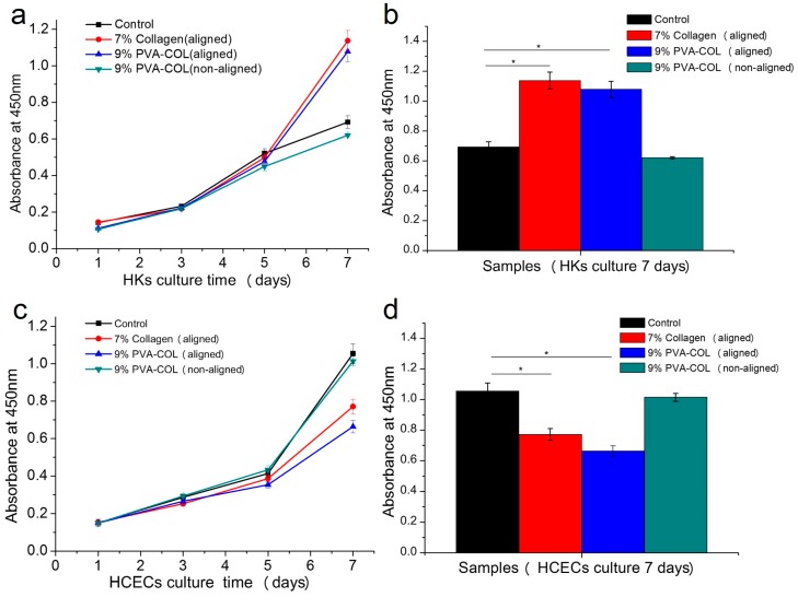
Corneal diseases are the main reason of vision loss globally. Constructing a corneal equivalent which has a similar strength and transparency with the native cornea, seems to be a feasible way to solve the shortage of donated cornea. Electrospun collagen scaffolds are often fabricated and used as a tissue-engineered cornea, but the main drawback of poor mechanical properties make it unable to meet the requirement for surgery suture, which limits its clinical applications to a large extent. Aligned polyvinyl acetate (PVA)/collagen (PVA-COL) scaffolds were electrospun by mixing collagen and PVA to reinforce the mechanical strength of the collagen electrospun scaffold. Human keratocytes (HKs) and human corneal epithelial cells (HCECs) inoculated on aligned and random PVA-COL electrospun scaffolds adhered and proliferated well, and the aligned nanofibers induced orderly HK growth, indicating that the designed PVA-COL composite nanofibrous electrospun scaffold is suitable for application in tissue-engineered cornea.

Keywords: corneal tissue; electrospun; nanofibrous scaffold.


Conflict of interest statement

The authors declare no conflict of interest.

Figures

Figure 1
Figure 1
SEM images of the non-aligned and aligned electrospun nanofibers, the diameter range and the alignment degree histograms of the respective electrospun scaffolds. Scale bar, 2 μm.
Figure 2
Figure 2
Mechanical properties and transparency test results: (a) average strain–stress curves of the 9% PVA-COL (aligned and non-aligned), 7% aligned PVA-COL and collagen electrospun scaffolds; (b) maximum tensile stress histogram of the samples; (c) the average transmittance of the 10% aligned PVA, 7% aligned collagen, 7% aligned PVA-COL and 9% PVA-COL (aligned and non-aligned) electrospun scaffolds under the wavelengths of 405, 450, 492 and 630 nm; and (d) transmittance histogram of the samples under the wavelength of 630 nm. The data represent the means ± SD (* p < 0.05).
Figure 3
Figure 3
The proliferation results of the HKs and HCECs: (a) proliferation of HKs on the 7% aligned collagen and the 9% PVA-COL (aligned and non-aligned) electrospun scaffolds within seven days; (b) proliferation histogram of the HKs in the different electrospun scaffolds cultured for seven days; (c) proliferation of HCECs on the 7% aligned collagen and the 9% PVA-COL (aligned and non-aligned) electrospun scaffolds within seven days; and (d) proliferation histogram of the HCECs in the different electrospun scaffolds cultured for seven days. The control group was the cells that were seeded on the culture plate, the data represent the means ± SD (* p < 0.05).
Figure 4
Figure 4
RFP labeled HKs: on the 7% aligned collagen (a,d,g); and the 9% PVA-COL aligned (b,e,h); and non-aligned (c,f,i) scaffolds that were cultured for: one week (ac); two weeks (df); and three weeks (gi). The scale bar is 500 μm.
Figure 5
Figure 5
The HKs growth on the 9% PVA-COL scaffolds: (ac) on the aligned scaffolds that were cultured for one, two and three weeks; and (df) on the random scaffolds that were cultured for one, two and three weeks. The scale bar is 200 μm. (gj) The aligned 9% PVA-COL scaffolds after culturing for four weeks. Scale bar: (g,h) 500 μm; (i) 200 μm; and (j) 100 μm.
Figure 6
Figure 6
The GFP labeled HCECs on the: aligned (ad); and non-aligned (eh) 9% PVA-COL scaffolds after culturing for: one week (a,e); two weeks (b,f); three weeks (c,g); and four weeks (d,h). The scale bar is 500 μm.
Figure 7
Figure 7
The HCECs on the: aligned (a,b,e,f) and non-aligned (c,d,g,h) 9% PVA-COL scaffolds after culturing for: three weeks (ad); and four weeks (eh). Scale bar: (a,c,e,g) 500 μm; and (b,d,f,h) 200 μm.

References

    1. Wilson S.L., Wimpenny I., Ahearne M., Rauz S., Haj A.J.E., Yang Y. Chemical and Topographical Effects on Cell Differentiation and Matrix Elasticity in a Corneal Stromal Layer Model. Adv. Funct. Mater. 2012;22:3641–3649. doi: 10.1002/adfm.201200655. - DOI
    1. Mclaughlin C.R., Tsai R.J., Latorre M.A., Griffith M. Bioengineered corneas for transplantation and in vitro toxicology. Front. Biosci. 2009;14:3326–3337. doi: 10.2741/3455. - DOI - PubMed
    1. Kong B., Mi S. Electrospun Scaffolds for Corneal Tissue Engineering: A Review. Materials. 2016;9:614. doi: 10.3390/ma9080614. - DOI - PMC - PubMed
    1. World Health Organization . Draft Action Plan for the Prevention of Avoidable Blindness and Visual Impairment 2014–2019: Towards Universal Eye Health: A Global Action Plan 2014–2019. Sixty-Sixth World Health Assembly; Geneva, Switzerland: 2013. Report by the Secretariat.
    1. Upadhyay M.P., Srinivasan M., Whitcher J.P. Corneal blindness: A global perspective. Bull. World Health Organ. 2001;79:214–221. - PMC - PubMed

LinkOut - more resources