. 2018 Feb 26;23(3):517.
doi: 10.3390/molecules23030517.

Aloin Suppresses Lipopolysaccharide-Induced Inflammatory Response and Apoptosis by Inhibiting the Activation of NF-κB

Affiliations

Aloin Suppresses Lipopolysaccharide-Induced Inflammatory Response and Apoptosis by Inhibiting the Activation of NF-κB

Xuan Luo et al. Molecules. .

Abstract

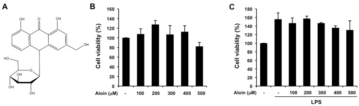
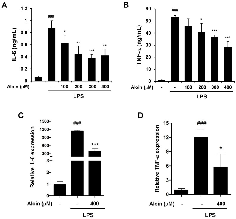
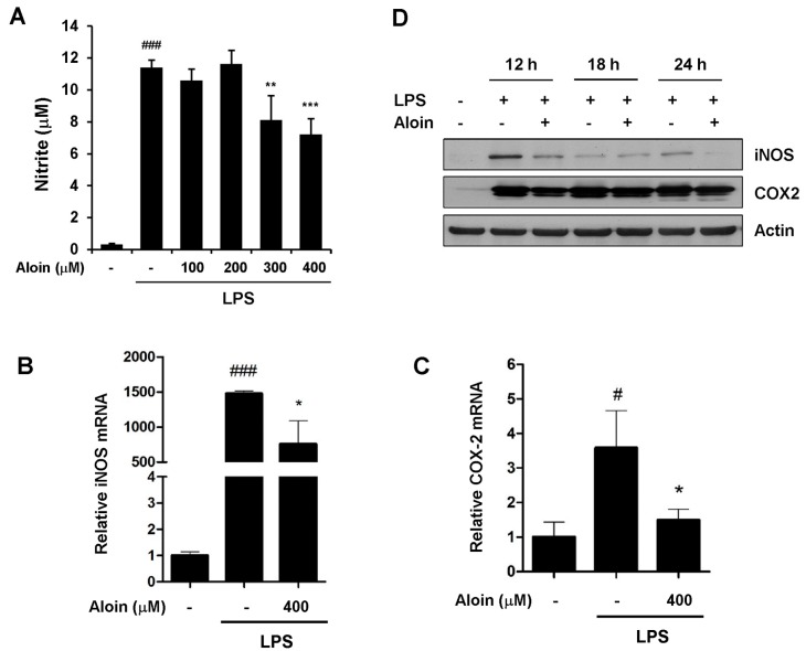
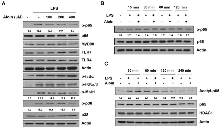
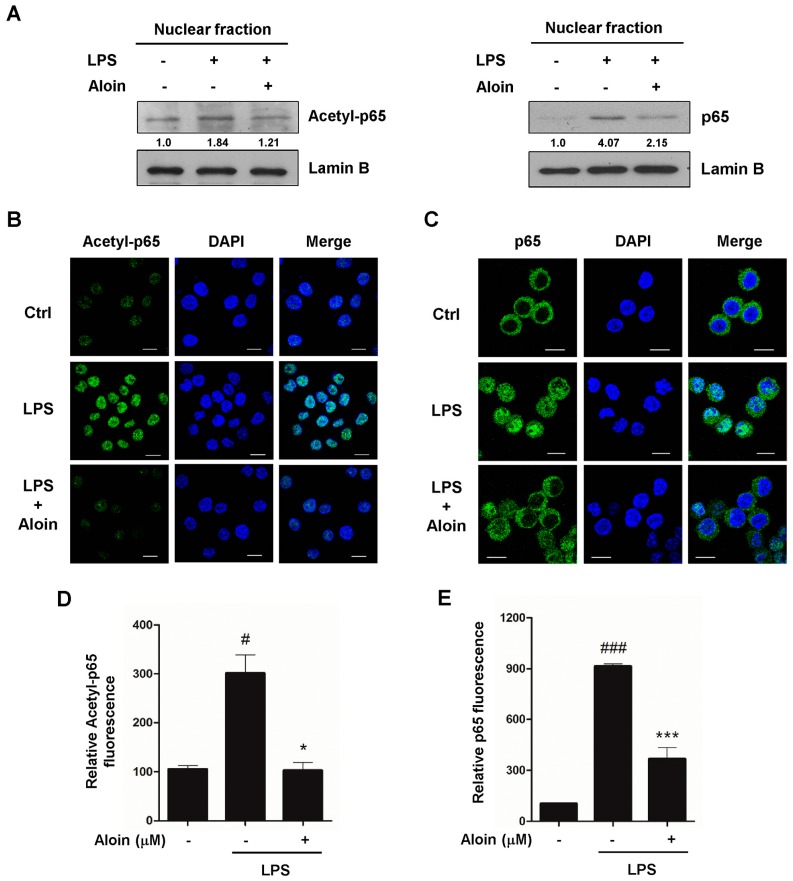
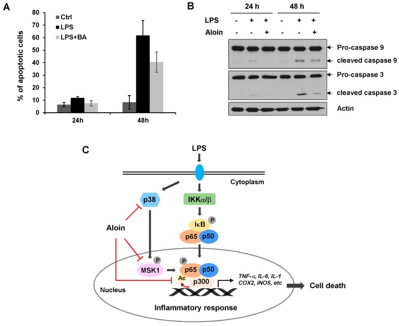
Numerous herbal-derived natural products are excellent anti-inflammatory agents. Several studies have reported that aloin, the major anthraquinone glycoside obtained from the Aloe species, exhibits anti-inflammatory activity. However, the molecular mechanism of this activity is not well understood. In this report, we found that aloin suppresses lipopolysaccharide-induced pro-inflammatory cytokine secretion and nitric oxide production, and downregulates the expression of tumor necrosis factor alpha (TNF-α), interleukin 6 (IL-6), inducible nitric oxide synthase (iNOS), and cyclooxygenase-2 (COX-2). Aloin inhibits the phosphorylation and acetylation of the NF-κB p65 subunit by suppressing the upstream kinases p38 and Msk1, preventing LPS-induced p65 translocation to the nucleus. We have also shown that aloin inhibits LPS-induced caspase-3 activation and apoptotic cell death. Collectively, these findings suggest that aloin effectively suppresses the inflammatory response, primarily through the inhibition of NF-κB signaling.

Keywords: NF-κB; aloin; apoptosis; inflammation; macrophages.


Conflict of interest statement

The authors declare no conflict of interest. The funding sponsors had no role in the design of the study; in the collection, analyses, or interpretation of data; in the writing of the manuscript, and in the decision to publish the results.

Figures

Figure 1
Figure 1
Aloin does not affect cell growth in murine macrophages: (A) Chemical structure of aloin; (B) RAW 264.7 cells were treated with various concentrations of aloin (100 μM, 200 μM, 300 μM, 400 μM, and 500 μM) for 24 h. The total number of viable cells was determined by methyl thiazolyl tetrazolium (MTT) assay. Values are mean ± SD of three independent experiments; (C) RAW 264.7 cells were treated with various concentrations of aloin and lipopolysaccharide (LPS) (100 ng/mL) for 24 h. The total number of viable cells was calculated. Values ARE mean ± SD of three independent experiments.
Figure 2
Figure 2
Aloin inhibits the LPS-induced expression of IL-6 and TNF-α RAW 264.7 cells were pre-treated with aloin for 2 h prior to LPS (100 ng/mL) stimulation. Cell-free supernatants were collected to detect (A) Interleukin 6 (IL-6) and (B) Tumor necrosis factor alpha (TNF-α) concentrations via ELISA after LPS treatment for 9 h and 24 h, respectively. The mRNA expression levels of (C) IL-6 and (D) TNF-α were determined by qPCR in RAW264.7 cells pre-treated with aloin (400 μM) followed by LPS (100 ng/mL) stimulation for 6 h. The data shown are the means ± SD of three experiments. ### p < 0.001 is significantly different from the control. * p < 0.05, ** p < 0.01, and *** p < 0.001 are different from the LPS alone.
Figure 3
Figure 3
Aloin inhibits nitric oxide (NO) production and the expression of cyclooxygenase-2 (COX-2) and inducible nitric oxide synthase (iNOS). (A) RAW 264.7 cells were pre-treated with aloin for 2 h prior to LPS (100 ng/mL) treatment. Cell-free supernatants were collected to determine NO production using the Griess reaction after LPS treatment for 24 h. The mRNA levels of (B) iNOS and (C) COX2 were determined by qPCR; (D) RAW 264.7 cells were pre-treated with or without 400 μM aloin for 2 h, and then stimulated with LPS (100 ng/mL) for 12 h, 18 h and 24 h. Levels of iNOS and COX-2 proteins were determined by Western blotting. The results shown are the means ± SD of three experiments. #p < 0.05 and ### p < 0.001 are significantly different from the control. * p < 0.05, ** p < 0.01, and *** p < 0.001 are different from the LPS alone.
Figure 4
Figure 4
Aloin inhibits LPS-induced NF-κB p65 phosphorylation and acetylation. (A) RAW 264.7 cells were pre-treated with various concentrations of aloin for 2 h, and stimulated with LPS (100 ng/mL) for 2 h. The protein expression levels of phospho-p65, total p65, MyD88, TLR4, TLR7, phosphor-IκBα, phospho-IKKα/β, phospho-Msk1, phospho-p38 and total p38 was determined by Western blotting; (B) Cells were pre-treated with or without aloin (400 μM) for 2 h, and stimulated with LPS (100 ng/mL) for 15 min, 30 min, 60 min, or 120 min. The protein levels of phospho-p65 and total p65 were analyzed by Western blotting; (C) Immunoblotting analysis of acetylated p65, total p65, and HDAC1 in RAW 264.7 cells treated with aloin and LPS at indicated time points.
Figure 5
Figure 5
Aloin inhibits LPS-induced NF-κB p65 nuclear translocation. (A) RAW 264.7 cells were pre-treated with or without aloin (400 μM) for 2 h, and then stimulated with LPS (100 ng/mL) for 2 h. Protein levels of acetylated p65 and total p65 in the nuclear fractions were analyzed by immunoblotting. Lamin B was used as a marker for nuclear fraction. RAW 264.7 cells treated with or without aloin and LPS were fixed and immunostained with (B) acetylated p65 or (C) total p65 antibodies, and fluorescent images were captured by confocal microscopy. Scale bar, 10 μm. At least 200 cells were counted for each sample; (D) Quantification of acetyl-p65 and (E) total p65 fluorescent intensities in RAW 264.7 cells. Data shown are the means ± SD of three experiments. # p < 0.05 and ### p < 0.001 are different from the control. * p < 0.05 and *** p < 0.001 are different from the LPS alone.
Figure 6
Figure 6
Aloin inhibits LPS-induced apoptotic cell death. (A) RAW 264.7 cells were pre-treated with or without aloin (400 μM) for 2 h, and then stimulated with LPS (100 ng/mL) for 24 h or 48 h. Cells were collected and stained with Annexin V/PI, the percentage of Annexin-V positive cells was analyzed by flow cytometry analysis Data shown are the means ± SD of three experiments; (B) The protein expression levels of pro-caspase 9, cleaved caspase-9, pro-caspase 3, and cleaved caspase 3 were determined by immunoblotting analysis. β-actin was used as a loading control; (C) Aloin prevented the LPS-induced inflammatory response by inhibiting NF-κB signaling. Aloin attenuated LPS-induced p65 post-translational modifications by inhibiting p38 and MSK1-mediated phosphorylation and p300-mediated acetylation. This in turn prevented p65 nuclear translocation, and downregulated NF-κB mediated gene expression, including pro-inflammatory cytokines, and genes involved in apoptosis.

References

    1. Oeckinghaus A., Ghosh S. The NF-kappaB Family of Transcription Factors and Its Regulation. Cold Spring Harb. Perspect. Biol. 2009;1:a000034. doi: 10.1101/cshperspect.a000034. - DOI - PMC - PubMed
    1. Tan A.C., Konczak I., Sze D.M., Ramzan I. Molecular Pathways for Cancer Chemoprevention by Dietary Phytochemicals. Nutr. Cancer. 2011;63:495–505. doi: 10.1080/01635581.2011.538953. - DOI - PubMed
    1. Tak P.P., Firestein G.S. NF-kappaB: A Key Role in Inflammatory Diseases. J. Clin. Investig. 2001;107:7–11. doi: 10.1172/JCI11830. - DOI - PMC - PubMed
    1. Neurath M.F., Pettersson S., Meyer zum Buschenfelde K.H., Strober W. Local Administration of Antisense Phosphorothioate Oligonucleotides to the p65 Subunit of NF-kappa B Abrogates Established Experimental Colitis in Mice. Nat. Med. 1996;2:998–1004. doi: 10.1038/nm0996-998. - DOI - PubMed
    1. Tomita T., Takeuchi E., Tomita N., Morishita R., Kaneko M., Yamamoto K., Nakase T., Seki H., Kato K., Kaneda Y., et al. Suppressed Severity of Collagen-Induced Arthritis by in Vivo Transfection of Nuclear Factor kappaB Decoy Oligodeoxynucleotides as a Gene Therapy. Arthritis Rheumatol. 1999;42:2532–2542. doi: 10.1002/1529-0131(199912)42:12<2532::AID-ANR5>3.0.CO;2-2. - DOI - PubMed

MeSH terms