Editorial
. 2018 Feb 27;11(1):27.
doi: 10.3390/ph11010027.

Glycosaminoglycans and Proteoglycans

Affiliations
Editorial

Glycosaminoglycans and Proteoglycans

Vitor H Pomin et al. Pharmaceuticals (Basel). .

Abstract

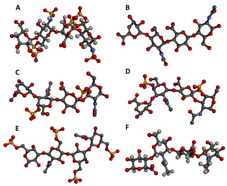
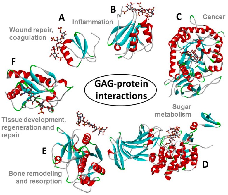
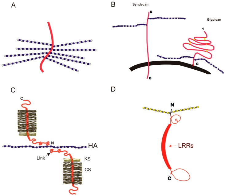
In this editorial to MDPI Pharmaceuticals special issue "Glycosaminoglycans and Proteoglycans" we describe in outline the common structural features of glycosaminoglycans and the characteristics of proteoglycans, including the intracellular proteoglycan, serglycin, cell-surface proteoglycans, like syndecans and glypicans, and the extracellular matrix proteoglycans, like aggrecan, perlecan, and small leucine-rich proteoglycans. The context in which the pharmaceutical uses of glycosaminoglycans and proteoglycans are presented in this special issue is given at the very end.

Keywords: chondroitin sulfate; decorin; dermatan sulfate; glycosaminoglycans; glypican; heparan sulfate; heparin; hyaluronan; keratan sulfate; perlecan; proteoglycans; serglycin; syndecan.


Conflict of interest statement

The authors declare no conflict of interest.

Figures

Figure 1
Figure 1
Three-dimensional tetrasaccharide representations, taken from files in the PDB as indicated, of: (A) Heparin [IdoA2S(α1→4)GlcNS6S(α1→4)IdoA2S(α1→4)GlcNS6S] from 1HPN; (B) heparan sulfate [GlcA(β1→4)GlcNAc(α1→4)GlcA(β1→4)GlcNAc] from 3E7J; (C) chondroitin 4-sulfate [GlcA(β1→3)GalNAc4S(β1→4)GlcA(β1→3)GalNAc4S] from 1OFM; (D) dermatan sulfate [IdoA(α1→3)GalNAc4S(β1→4)IdoA(α1→3)GalNAc4S] from 1OFL; (E) keratan sulfate [Gal6S(β1→4)GlcNAc6S(β1→3)Gal6S(β1→4)GlcNAc6S] from 1KES; and (F) hyaluronan [GlcA(β1→3)GlcNAc(β1→4)GlcA(β1→3)GlcNAc] from 2BVK. The atoms in the ball-stick representations are carbon (grey), nitrogen (blue), hydrogen (light grey); oxygen (red) and sulfur (yellow). A and F are NMR solution structures, and non-exchangeable protons are shown; the others are crystal structures so are shown without protons.
Figure 2
Figure 2
Structural representations from the crystal structures of some illustrative GAG-protein complexes in PDB: (A) macrophage inflammatory protein 1-alpha (CCL3) monomer + Hp 4-mer (from 5D65); (B) platelet factor 4 (CXCL4) dimer + fondaparinux (antithrombin-high affinity Hp 5-mer) (from 4R9W); (C) human heparanase complex + Hp tetrasaccharide (from 5E9C); (D) d-glucuronyl C5-epimerase + Hp 6-mer (from 4PXQ); (E) cathepsin K monomer + DS 6-mer (from 4N79); and (F) Sonic Hedgehog (Shh) monomer + CS-A 4-mer (from 4C4M). The atoms of the GAG ligands represented in the ball-stick view are carbon (grey), nitrogen (blue), hydrogen (light grey); oxygen (red) and sulfur (yellow). In the proteins, the alpha-helices, beta-sheets, loops, and random coils are represented, respectively, in red, blue, green and grey. The pathophysiological systems in which these complexes play a role are indicated by grey fonts in the panel.
Figure 3
Figure 3
Schematic cartoon images (not to scale) for PGs on the cell surface and in the ECM. Protein chains are shown as red ribbons, and GAG chains are depicted in a simplified form of the symbology recommended by the Consortium for Structural Glycomics [40]; (A) the intracellular PG serglycin, bearing closely packed Hp (or oversulfated chondroitin) chains, on a small peptide core; (B) cell-surface PGs syndecan and glypican; the cell membrane is shown in black; (C) the complex between aggrecan and HA, mediated by Link protein, that forms the structural basis for cartilage elasticity; and (D) a generic diagram of a SLRP, such as biglycan or decorin. Between the globular regions near the N- and C-termini the leucine-rich repeats (LRRs) form a curved structure; the dimers can form by interaction between the two offset concave faces of monomers.

References

    1. Mulloy B., Hogwood J., Gray E., Lever R., Page C.P. Pharmacology of Heparin and Related Drugs. Pharmacol. Rev. 2016;68:76–141. doi: 10.1124/pr.115.011247. - DOI - PubMed
    1. Mantovani V., Maccari F., Volpi N. Chondroitin Sulfate and Glucosamine as Disease Modifying Anti-Osteoarthritis Drugs (DMOADs) Curr. Med. Chem. 2016;23:1139–1151. doi: 10.2174/0929867323666160316123749. - DOI - PubMed
    1. Weyers A., Linhardt R.J. Neoproteoglycans in tissue engineering. FEBS J. 2013;280:2511–2522. doi: 10.1111/febs.12187. - DOI - PMC - PubMed
    1. Sasisekharan R., Venkataraman G. Heparin and heparan sulfate: Biosynthesis, structure and function. Curr. Opin. Chem. Biol. 2000;4:626–631. doi: 10.1016/S1367-5931(00)00145-9. - DOI - PubMed
    1. Rabenstein D.L. Heparin and heparan sulfate: Structure and function. Nat. Prod. Rep. 2002;19:312–331. doi: 10.1039/b100916h. - DOI - PubMed

Publication types

LinkOut - more resources