Review
. 2018 Feb 27;10(1):28.
doi: 10.3390/pharmaceutics10010028.

Ocular Drug Delivery Barriers-Role of Nanocarriers in the Treatment of Anterior Segment Ocular Diseases

Affiliations
Review

Ocular Drug Delivery Barriers-Role of Nanocarriers in the Treatment of Anterior Segment Ocular Diseases

Rinda Devi Bachu et al. Pharmaceutics. .

Abstract

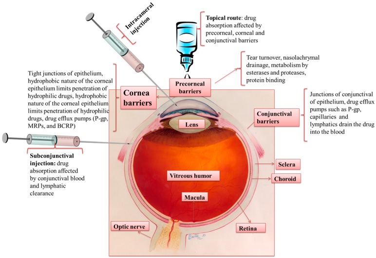
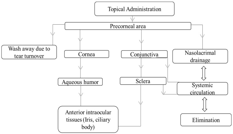
Ocular drug delivery is challenging due to the presence of anatomical and physiological barriers. These barriers can affect drug entry into the eye following multiple routes of administration (e.g., topical, systemic, and injectable). Topical administration in the form of eye drops is preferred for treating anterior segment diseases, as it is convenient and provides local delivery of drugs. Major concerns with topical delivery include poor drug absorption and low bioavailability. To improve the bioavailability of topically administered drugs, novel drug delivery systems are being investigated. Nanocarrier delivery systems demonstrate enhanced drug permeation and prolonged drug release. This review provides an overview of ocular barriers to anterior segment delivery, along with ways to overcome these barriers using nanocarrier systems. The disposition of nanocarriers following topical administration, their safety, toxicity and clinical trials involving nanocarrier systems are also discussed.

Keywords: anterior segment; disposition; novel drug delivery systems; polymeric nanocarriers; toxicity.


Conflict of interest statement

The authors declare no conflict of interest.

Figures

Figure 1
Figure 1
Routes of administration for anterior segment drug delivery, ocular tissue barriers and clearance mechanisms that prevent drug absorption into the eye. Modified from: Credit: National Eye Institute, National Institutes of Health (Ref#: NEA04).
Figure 2
Figure 2
Concentration of cyclosporine A (CsA) in the cornea after the instillation of formulations (* p < 0.05) Reprinted from [59]. Copyright (2018), with permission from Elsevier.
Figure 3
Figure 3
Anti-bacterial activity of optimized moxifloxacin-loaded nanosuspension (MN4) vs. marketed eye drops (M®) [68].
Figure 4
Figure 4
Concentration of ciprofloxacin in rabbit aqueous humor after the instillation of various liposomal formulations (F6, F12, and F13) as well as commercial eye drops. F6, F12, F13 are prepared using varying molar ratios of phosphatidylcholine (PC), Dipalmitoylphosphatidylcholine (DPPC), 1,2-Dimyristoyl-sn-glycero-3-phosphocholine (DMPC), cholesterol (CH), stearylamine (SA) and dioctadecyldimethyl ammonium bromide (DODAB). Reprinted from [74]. Copyright (2018), with permission from Elsevier.
Figure 5
Figure 5
Change in intraocular pressure (IOP) for various formulations in (a) dosed eye (b) contralateral eye (n = 6). (TMS—Timolol solution 0.25%, REVTM—Uncoated niosomes, REVTMbio1-chitosan coated niosomes, REVTMbio2 and 3-carbopol coated niosomes, GFS—Timolet® GFS). Reprinted from [86]. Copyright (2018), with permission from Elsevier.
Figure 6
Figure 6
Concentration-time profile of Dexamethasone (Dex) in the aqueous humor of rabbit after the instillation of various formulations. Reprinted from [89]. Copyright (2018), with permission from Elsevier.
Figure 7
Figure 7
Cell viability of human corneal epithelial cells (HCE) when treated with different concentrations of blank (white columns) and cyclosporine A-loaded micelles (dark columns). Reprinted from [107]. Copyright (2018), with permission from Elsevier.
Figure 8
Figure 8
Concentration-time profile of Tobramycin (TOB) in the aqueous humor of rabbit after the instillation of the reference solution (■) and TOB-SLN (♦). Reprinted from [147]. Copyright (2018), with permission from Elsevier.
Figure 9
Figure 9
In vivo real time fluroscence imaging of timolol maleate (TM) eye drops, gel, liposomes and composite system (TM L-ISG). Recovery of TM on the corneal surface at 5 s, 1.5, 3, 5 and 10 min post-application. Reprinted from [162]. Copyright (2018), with permission from Elsevier.
Figure 10
Figure 10
Possible disposition routes of nanocarriers following topical administration.

References

    1. Patel A., Cholkar K., Agrahari V., Mitra A.K. Ocular drug delivery systems: An overview. World J. Pharmacol. 2013;2:47–64. doi: 10.5497/wjp.v2.i2.47. - DOI - PMC - PubMed
    1. Boddu S.H.S., Menees A.L., Ray A., Mitra A.K. A Brief Overview of Ocular Anatomy and Physiology. In: Mitra A.K., editor. Treatise on Ocular Drug Delivery. Bentham Science Publishers; Sharjah, UAE: 2013. pp. 3–19.
    1. Pascolini D., Mariotti S.P. Global estimates of visual impairment: 2010. Bri. J. Ophthalmol. 2012;96:614–618. doi: 10.1136/bjophthalmol-2011-300539. - DOI - PubMed
    1. Boddu S.H.S., Gunda S., Earla R., Mitra A.K. Ocular microdialysis: A continuous sampling technique to study pharmacokinetics and pharmacodynamics in the eye. Bioanalysis. 2010;2:487–507. doi: 10.4155/bio.10.2. - DOI - PubMed
    1. Klyce S.D., Crosson C.E. Transport processes across the rabbit corneal epithelium: A review. Curr. Eye Res. 1985;4:323–331. doi: 10.3109/02713688509025145. - DOI - PubMed

LinkOut - more resources