. 2018 Feb 28;19(3):684.
doi: 10.3390/ijms19030684.

Loss of BID Delays FASL-Induced Cell Death of Mouse Neutrophils and Aggravates DSS-Induced Weight Loss

Affiliations

Loss of BID Delays FASL-Induced Cell Death of Mouse Neutrophils and Aggravates DSS-Induced Weight Loss

Simone Wicki et al. Int J Mol Sci. .

Abstract

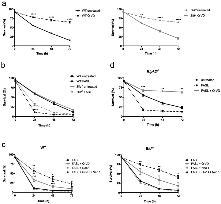
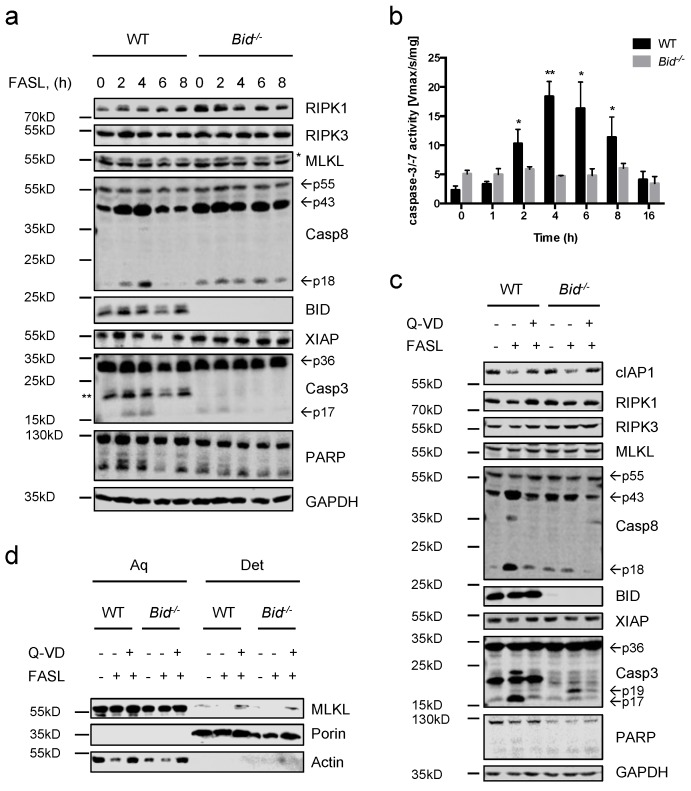
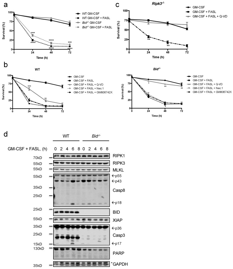
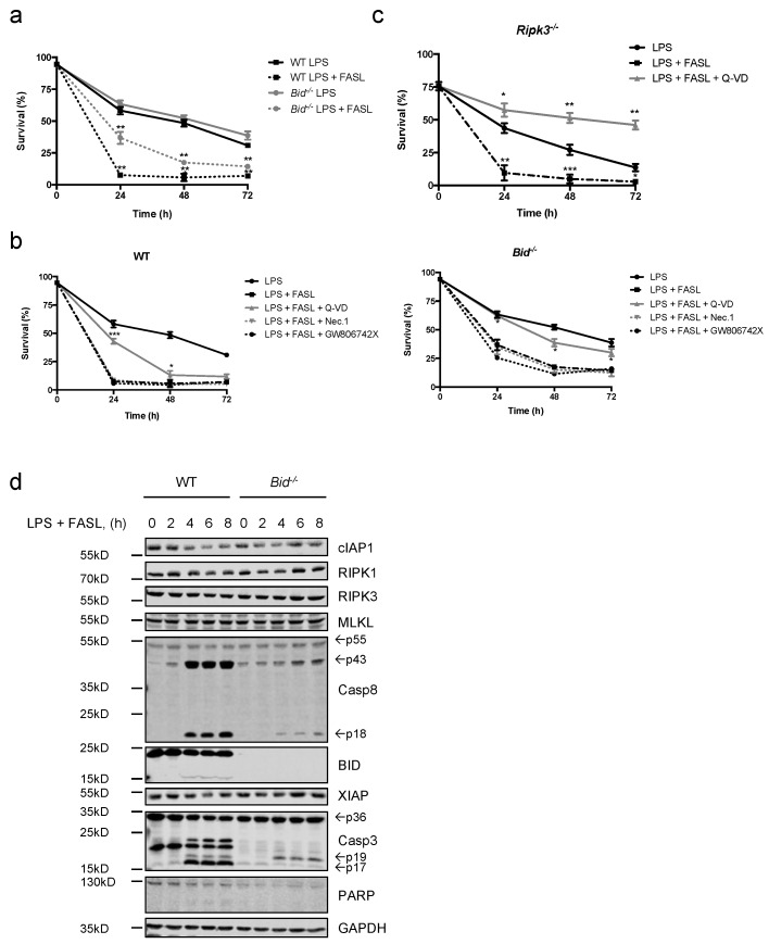
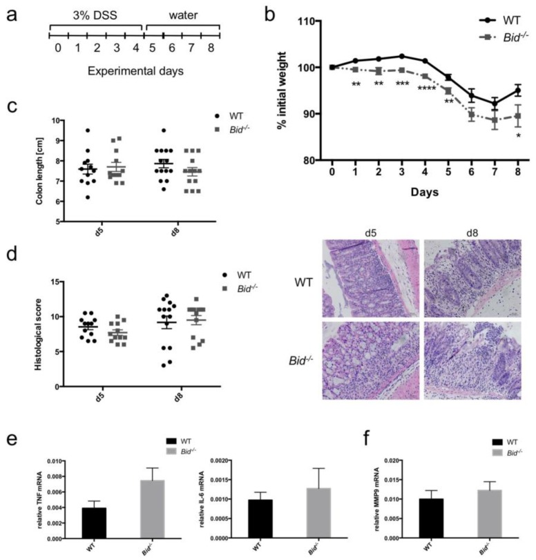
Neutrophils are key players in the early defense against invading pathogens. Due to their potent effector functions, programmed cell death of activated neutrophils has to be tightly controlled; however, its underlying mechanisms remain unclear. Fas ligand (FASL/CD95L) has been shown to induce neutrophil apoptosis, which is accelerated by the processing of the BH3-only protein BH3 interacting domain death agonist (BID) to trigger mitochondrial apoptotic events, and been attributed a regulatory role during viral and bacterial infections. Here, we show that, in accordance with previous works, mouse neutrophils underwent caspase-dependent apoptosis in response to FASL, and that this cell death was significantly delayed upon loss of BID. However, pan-caspase inhibition failed to protect mouse neutrophils from FASL-induced apoptosis and caused a switch to RIPK3-dependent necroptotic cell death. Intriguingly, such a switch was less evident in the absence of BID, particularly under inflammatory conditions. Delayed neutrophil apoptosis has been implicated in several auto-inflammatory diseases, including inflammatory bowel disease. We show that neutrophil and macrophage driven acute dextran sulfate sodium (DSS) induced colitis was slightly more aggravated in BID-deficient mice, based on significantly increased weight loss compared to wild-type controls. Taken together, our data support a central role for FASL > FAS and BID in mouse neutrophil cell death and further underline the anti-inflammatory role of BID.

Keywords: BID; FAS/CD95; RIPK3; apoptosis; caspases; colitis; inflammation; mouse model; necroptosis; neutrophil.


Conflict of interest statement

The authors declare no conflict of interest.

Figures

Figure 1
Figure 1
Mouse neutrophils undergo cell death in response to FASL, which is delayed by loss of BID. (a) Assessment of viability of WT and Bid−/− neutrophils left untreated (spontaneous apoptosis) or treated with the pan-caspase inhibitor Q-VD-OPh (20 μM) for indicated time points. n ≥ 6, mean ± SEM. (b) Assessment of viability of WT and Bid−/− neutrophils upon treatment with FASL (100 ng/mL) for indicated time points. n ≥ 3, mean ± SEM. (c) WT and Bid−/− neutrophils were pre-treated with Q-VD-OPh (20 μM) and/or Nec.1 (20 μM) for 30 min followed by treatment with FASL (100 ng/mL) for indicated time points. Viability was assessed by flow cytometry. n ≥ 4, mean ± SEM. (d) Ripk3−/− neutrophils were pre-treated with Q-VD-OPh (20 μM) for 30 min and subsequently stimulated with FASL (100 ng/mL) for indicated time points. Viability was assessed by flow cytometry using GFP-AnnexinV/PI exclusion. n = 3, mean ± SEM. All experiments were performed with primary bone marrow-derived neutrophils. p < 0.05 (*), p < 0.01 (**), p < 0.005 (***) and p < 0.001 (****).
Figure 2
Figure 2
FASL induces apoptosis in neutrophils, which switches to necroptosis when caspases are inhibited. (a) WT and Bid−/− neutrophils were treated with FASL (100 ng/mL) for 0–8 h. Lysates were assessed by immunoblotting. Presented immunoblots are representative of at least two independent experiments. * indicates non-specific bands; ** indicates bands derived from previous anti-BID immunoblotting. (b) WT and Bid−/− neutrophils were stimulated with FASL (100 ng/mL) for indicated time points. Lysates were assayed for caspase-3/-7 activity by fluorogenic DEVDase assay. n ≥ 3, mean ± SEM. p < 0.05 (*), p < 0.01 (**). (c) WT and Bid−/− neutrophils were pre-treated with Q-VD-OPh (20 μM) for 30 min and subsequently treated with FASL (100 ng/mL) for 6 h. Lysates were subjected to immunoblotting. Presented immunoblots are representative of at least two independent experiments. (d) WT and Bid−/− neutrophils were pre-stimulated with Q-VD-OPh (20 μM) for 30 min followed by administration of FASL (100 ng/mL) for 16 h. Fractionation by phase separation was performed and lysates were subjected to immunoblotting. Presented immunoblots are representative of at least two independent experiments. All experiments were performed with in vitro differentiated neutrophils.
Figure 3
Figure 3
GM-CSF-primed neutrophils remain sensitive to FASL killing. (a) Primary WT and Bid−/− neutrophils were primed with GM-CSF (1 ng/mL) for 30 min prior to stimulation with FASL (100 ng/mL) for indicated time points. Viability was assessed by flow cytometry. n ≥ 3, mean ± SEM. (b) Primary WT and Bid−/− neutrophils were primed with GM-CSF (1 ng/mL) for 30 min and pre-treated with either Q-VD-OPh (20 μM), Nec.1 (20 μM) or the mouse MLKL inhibitor GW806742X (1 μM) for 30 min followed by stimulation with FASL (100 ng/mL) for indicated time points. Viability was assessed by flow cytometry. n ≥ 3, mean ± SEM. (c) In vitro differentiated Ripk3−/− neutrophils were primed with GM-CSF (1 ng/mL) for 30 min prior to stimulation with FASL (100 ng/mL) with or without Q-VD-OPh (20 μM) for indicated time points. Viability was assessed by flow cytometry. n ≥ 3, mean ± SEM. (ac): p < 0.05 (*), p < 0.01 (**), p < 0.005 (***) and p < 0.001 (****). (d) In vitro differentiated WT and Bid−/− neutrophils were primed with GM-CSF (1 ng/mL) for 30 min and subsequently treated with FASL (100 ng/mL) for 0–8 h. Lysates were assessed by immunoblotting. Presented immunoblots are representative of at least two independent experiments.
Figure 4
Figure 4
LPS-primed neutrophils remain sensitive to FASL killing. (a) Primary WT and Bid−/− neutrophils were primed with LPS (10 ng/mL) for 30 min prior to stimulation with FASL (100 ng/mL) for indicated time points. Viability was assessed by flow cytometry. n ≥ 3, mean ± SEM. (b) Primary WT and Bid−/− neutrophils were primed with LPS (10 ng/mL) for 30 min, pre-treated with either Q-VD-OPh (20 μM), Nec.1 (20 μM) or the mouse MLKL inhibitor GW806742X (1 μM) for 30 min followed by stimulation with FASL (100 ng/mL) for indicated time points. Viability was assessed by flow cytometry. n = 3, mean ± SEM. (c) In vitro differentiated Ripk3−/− neutrophils were primed with LPS (10 ng/mL) for 30 min prior to stimulation with FASL (100 ng/mL) with or without Q-VD-OPh (20 μM) for indicated time points. Viability was assessed by flow cytometry. n ≥ 3, mean ± SEM. (ac): p < 0.05 (*), p < 0.01 (**) and p < 0.005 (***). (d) In vitro differentiated WT and Bid−/− neutrophils were primed with LPS (10 ng/mL) prior to stimulation with FASL (100 ng/mL) for 0–8 h. Lysates were subjected to immunoblot. Presented immunoblots are representative of at least two independent experiments.
Figure 5
Figure 5
DSS-induced colitis is mildly exacerbated upon loss of BID. Colitis induced by 3% DSS in WT and Bid−/− mice (n = 12–14 animals per time point and group). (a) Scheme of the experimental set up. (b) Time course of weight loss (p < 0.05 (*), p < 0.01 (**), p < 0.005 (***) and p < 0.001 (****)) and (c) colon length after Day 5 and Day 8 of treatment. (d) Histology of colon tissues stained with H&E after Day 5 and Day 8 of treatment and analysis of severity of colitis by histological scoring. Presented images are representative. (e) Relative Tnfα and Il-6 mRNA expression in colon tissues after five days of treatment. Gapdh was used as reference gene. (f) Relative Mmp9 mRNA expression in colon tissues after five days of treatment. Gapdh was used as reference gene.

References

    1. Borregaard N. Neutrophils, from marrow to microbes. Immunity. 2010;33:657–670. doi: 10.1016/j.immuni.2010.11.011. - DOI - PubMed
    1. Galani I.E., Andreakos E. Neutrophils in viral infections: Current concepts and caveats. J. Leukoc. Biol. 2015;98:557–564. doi: 10.1189/jlb.4VMR1114-555R. - DOI - PubMed
    1. Geering B., Stoeckle C., Conus S., Simon H.U. Living and dying for inflammation: Neutrophils, eosinophils, basophils. Trends Immunol. 2013;34:398–409. doi: 10.1016/j.it.2013.04.002. - DOI - PubMed
    1. Duffin R., Leitch A.E., Fox S., Haslett C., Rossi A.G. Targeting granulocyte apoptosis: Mechanisms, models, and therapies. Immunol. Rev. 2010;236:28–40. doi: 10.1111/j.1600-065X.2010.00922.x. - DOI - PubMed
    1. Colotta F., Re F., Polentarutti N., Sozzani S., Mantovani A. Modulation of granulocyte survival and programmed cell death by cytokines and bacterial products. Blood. 1992;80:2012–2020. - PubMed

MeSH terms