. 2018 May;38(5):1102-1114.
doi: 10.1161/ATVBAHA.118.310703. Epub 2018 Mar 1.

Ly6CHi Blood Monocyte/Macrophage Drive Chronic Inflammation and Impair Wound Healing in Diabetes Mellitus

Affiliations

Ly6CHi Blood Monocyte/Macrophage Drive Chronic Inflammation and Impair Wound Healing in Diabetes Mellitus

Andrew Kimball et al. Arterioscler Thromb Vasc Biol. 2018 May.

Abstract

Objective: Wound monocyte-derived macrophage plasticity controls the initiation and resolution of inflammation that is critical for proper healing, however, in diabetes mellitus, the resolution of inflammation fails to occur. In diabetic wounds, the kinetics of blood monocyte recruitment and the mechanisms that control in vivo monocyte/macrophage differentiation remain unknown.

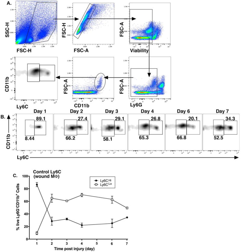
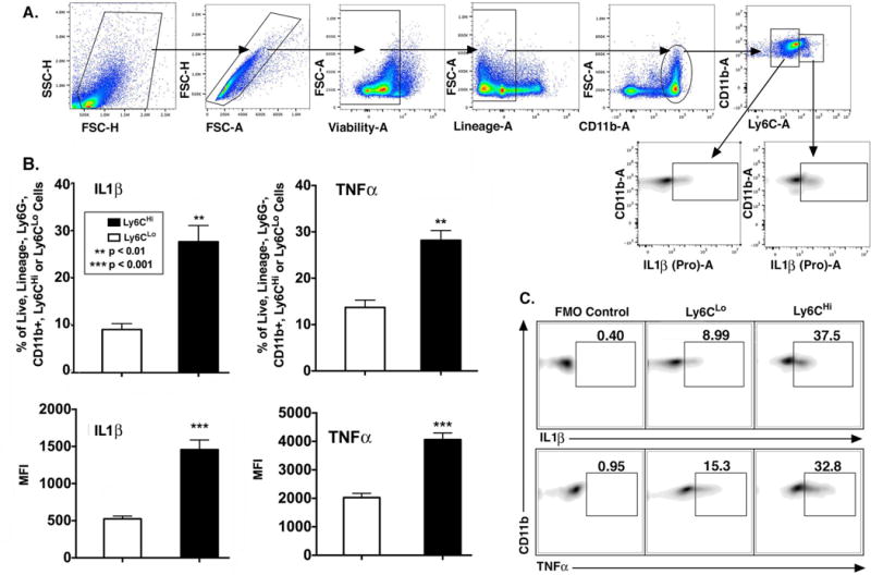
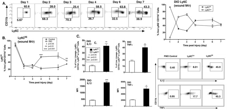
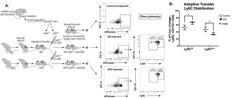
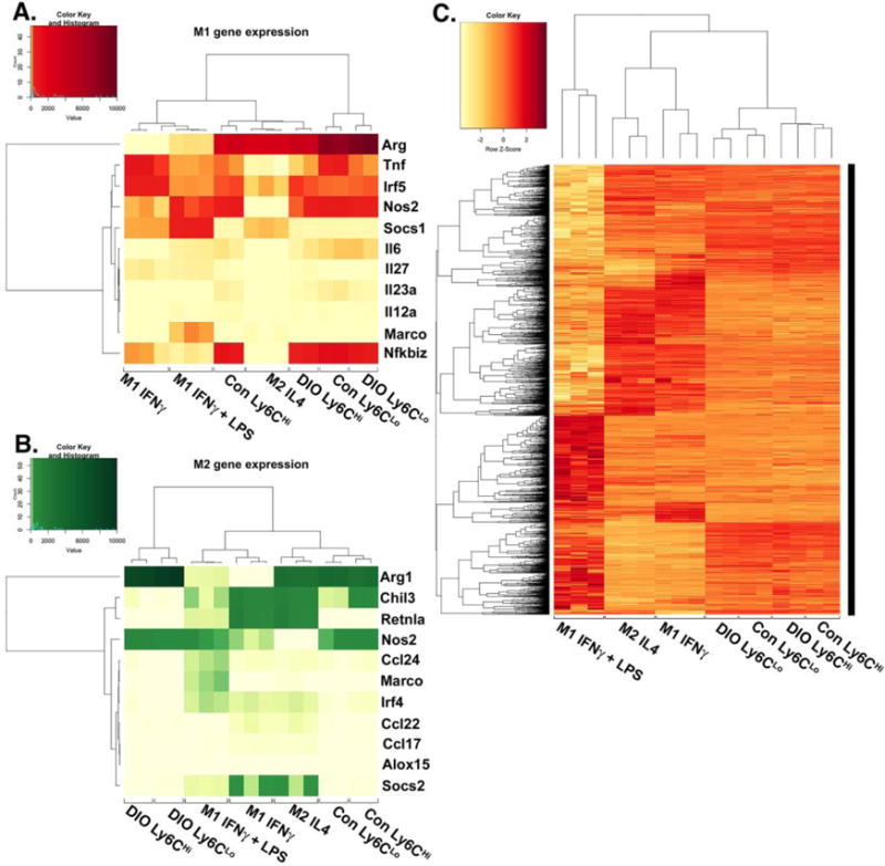
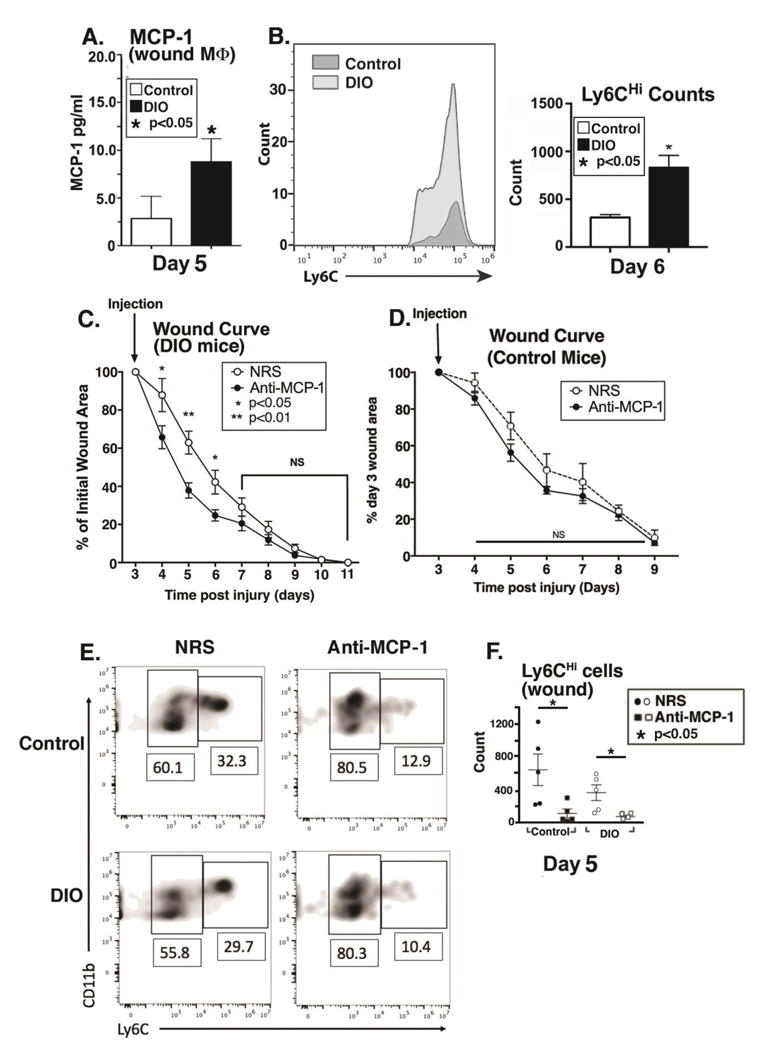
Approach and results: Here, we characterized the kinetics and function of Ly6CHi [Lin- (CD3-CD19-NK1.1-Ter-119-) Ly6G-CD11b+] and Ly6CLo [Lin- (CD3-CD19-NK1.1-Ter-119-) Ly6G-CD11b+] monocyte/macrophage subsets in normal and diabetic wounds. Using flow-sorted tdTomato-labeled Ly6CHi monocyte/macrophages, we show Ly6CHi cells transition to a Ly6CLo phenotype in normal wounds, whereas in diabetic wounds, there is a late, second influx of Ly6CHi cells that fail transition to Ly6CLo. The second wave of Ly6CHi cells in diabetic wounds corresponded to a spike in MCP-1 (monocyte chemoattractant protein-1) and selective administration of anti-MCP-1 reversed the second Ly6CHi influx and improved wound healing. To examine the in vivo phenotype of wound monocyte/macrophages, RNA-seq-based transcriptome profiling was performed on flow-sorted Ly6CHi [Lin-Ly6G-CD11b+] and Ly6CLo [Lin-Ly6G-CD11b+] cells from normal and diabetic wounds. Gene transcriptome profiling of diabetic wound Ly6CHi cells demonstrated differences in proinflammatory and profibrotic genes compared with controls.

Conclusions: Collectively, these data identify kinetic and functional differences in diabetic wound monocyte/macrophages and demonstrate that selective targeting of CD11b+Ly6CHi monocyte/macrophages is a viable therapeutic strategy for inflammation in diabetic wounds.

Keywords: diabetes mellitus; inflammation; macrophages; monocytes; wound healing.


Conflict of interest statement

Conflict of Interest Statement: The authors have no conflict of interest to declare.

Figures

Figure 1
Figure 1. Ly6CHi[Live,Ly6G,CD11b+] monocyte/macrophages rapidly accumulate during the first 24–48 hours following injury and then transition to Ly6CLo[Live,Ly6G,CD11b+] cells
Wounds were created using a 4mm punch biopsy on the back of male C57BL/6 mice. Wounds were harvested daily for 7 days post-injury and analyzed by flow cytometry. (A) Gating strategy to select single, live, Ly6G, CD11b+ cells and stratify by Ly6CHi vs. Ly6CLo. (B) Representative flow plots of Ly6CHi[Live,Ly6G,CD11b+] and Ly6CLo[Live,Ly6G,CD11b+] cells on post-injury days 1-7. (C) Ly6CHi[Live,Ly6G,CD11b+] and Ly6CLo[Live,Ly6G,CD11b+] cells plotted as a percentage of CD11b+ cells (n =32 mice; tissues of 2 wounds per mouse were pooled for a single biological replicate. Data is representative of 3 independent experiments.) All data are expressed as the mean +/− the standard error of the mean (SEM).
Figure 2
Figure 2. Wound CD11b+Ly6CHi and CD11b+Ly6CLo monocyte/macrophages display distinct inflammatory profiles
Wounds from C57BL/6 mice were collected on post-injury day 2 for cell isolation, ex vivo stimulation, and intra-cellular staining for flow cytometry. (A) Gating strategy to select single, live, lineage [CD3,CD19,NK1.1,Ter-119] , Ly6G, CD11b+, Ly6CHi and Ly6CLo cells. (B) Top panels: Percentage (%) of Ly6CHi vs. Ly6CLo cells staining positive for IL1β and TNFα. Bottom panels: MFI of IL1β and TNFα in the Ly6CHi and Ly6CLo gate (**P < 0.01, ***P < 0.001; n = 10 mice; tissues of 2 wounds per mouse were pooled for a single biological replicate. Data is representative of 3 independent experiments.) (C) Representative density plots of Ly6CHi and Ly6CLo cells stratified for IL1β and TNFα with FMO control. All data are expressed as mean +/− the standard error of the mean (SEM).
Figure 3
Figure 3. Diabetic wounds demonstrate a second, late influx of CD11b+Ly6CHi monocyte/macrophages
C57BL/6 mice were fed high fat (HFD) chow (60%kCal) for 12–16 weeks to induce obesity and insulin resistance/glucose intolerance in the diet-induced obese model (DIO) of physiologic type 2 diabetes. Wounds were collected on post-injury days 1–7 for flow cytometry. (A) Representative density plots of DIO Ly6CHi[Live,Ly6G,CD11b+] and DIO Ly6CLo[Live,Ly6G,CD11b+] cells on post-injury days 1-7. Gating strategy was identical to Figure 1. DIO Ly6CHi[Live, Ly6G,CD11b+] and DIO Ly6CLo[Live, Ly6G,CD11b+] cells plotted as a percentage of CD11b+ cells (n = 32 mice; tissues of 2 wounds per mouse were pooled for a single biological replicate. Data is representative of 3 independent experiments.) (B) Direct comparison of control and DIO Ly6CHi[Live,Ly6G,CD11b+] cells over time following injury. (*P < 0.05,**P < 0.01, ***P < 0.001; n = 4 mice/group/time point; tissues of 2 wounds per mouse were pooled for a single biological replicate. Data is representative of 2 experiments.) (C) DIO wounds were collected on post-injury day 2 for cell isolation, ex vivo stimulation, and intra-cellular staining for flow cytometry. Representative density plots of DIO Ly6CHi and Ly6CLo [Lin, Ly6G,CD11b+] cells as gated in Figure 2, stratified for IL1β and TNFα. Top panels: % of DIO CD11b+Ly6CHi vs. DIO CD11b+Ly6CLo cells staining positive for IL1β and TNFα. Bottom panels: MFI of IL1β and TNFα in the DIO CD11b+Ly6CHi and DIO CD11b+Ly6CLo gate (*P <0.05, **P < 0.01, ***P < 0.001; n = 5 mice/group; tissues of 2 wounds per mouse were pooled for a single biological replicate. Data is representative of 2 experiments.)
Figure 4
Figure 4. Diabetic wound CD11b+Ly6CHi cells demonstrate delayed transition to a CD11b+Ly6CLo phenotype
(A) Schematic of adoptive transfer experiment in which 106 tdTomato-expressing Ly6CHi[Lin, Ly6G, CD11b+] peripheral blood cells were injected via tail vein into control and DIO mice 24 hours after wounding. Control and DIO wounds were harvested on day 4 (3 days post-adoptive transfer) and single cell suspensions were processed for flow cytometry. Representative density plots of adoptively transferred control and DIO wounds gated for tdTomato+[LinLy6GCD11b+] cells and then stratifying by Ly6C designation. (B) Control and DIO wounds were harvested on day 4 (3 days post-adoptive transfer) and single cell suspensions were processed for flow cytometry. Relative percentages of adoptively transferred Ly6CHi[LinLy6GCD11b+tdTomato+] and Ly6CLo[LinLy6GCD11b+tdTomato+] monocyte/macrophages (*P < 0.05; n = 6 wounds per group, repeated 1X). All data are expressed as the mean +/− the standard error.
Figure 5
Figure 5. Diabetic wound CD11b+Ly6CHi and CD11b+Ly6CLo monocyte/macrophages display distinct transcriptome profiles
Wounds from normal and high fat diet (HFD)-fed, diet-induced obese (DIO) C57BL/6 mice were collected on post-injury day 3 for cell isolation, surface staining, and FACs to isolate Ly6CHi[LinLy6GCD11b+] and Ly6CLo[LinLy6GCD11b+] monocyte/macrophages for RNA-sequencing (RNA-seq). RNA samples were processed by the NIH-funded, University of Michigan DNA sequencing core. Reads were trimmed using Trimmomatic and mapped using HiSAT2. Read counts were performed using the featureCounts option from the subRead package followed by the elimination of low reads and normalization using edgeR. For comparison to in vitro generated macrophages, M1[IFNγ], M1[LPS,IFNγ] and M2[IL-4] datasets were used from publically available transcriptome data generated by Piccolo et al. (A) A heatmap of normalized reads obtained from edgeR for an internationally recognized panel of consensus genes for in vitro generated M1 macrophages. Wound Ly6CHi[LinLy6GCD11b+] and Ly6CLo[LinLy6GCD11b+] monocyte/macrophages were compared to the M1[IFNγ], M1[LPS,IFNγ], and M2[IL-4] transcriptomes. (B) A heatmap of normalized reads obtained from edgeR for an internationally recognized panel of consensus genes for in vitro generated M2 macrophages. Wound Ly6CHi[LinLy6GCD11b+] and Ly6CLo[LinLy6GCD11b+] monocyte/macrophages were compared to the M1[IFNγ], M1[LPS,IFNγ], and M2[IL-4] transcriptome. (C) Clustered Pearson correlation of the entire transcriptome of in vivo DIO and control Ly6CHi[LinLy6GCD11b+] and Ly6CLo[LinLy6GCD11b+] monocyte/macrophages and in vitro M1[IFNγ], M1[LPS,IFNγ], and M2[IL-4] macrophages (n = 8 mice per group; 4 wounds per mouse were pooled to obtain a single biological replicate given low RNA volumes. Two biological replicates per group were examined.)
Figure 6
Figure 6. Timed treatment of diabetic mice with anti-MCP-1 antibody post-injury restores normal healing
(A) The ligand for the blood monocyte CCR2 receptor, MCP-1, was examined in wound macrophages isolated by magnetic cell sorting CD11b+[CD3CD19NK1.1Ly6G] from DIO and control mice on day 5. Protein levels of MCP-1 as determined by ELISA in control and DIO wound cells on day 5 post-injury. (*P < 0.05; n= 5 mice/group; data is representative of 2 independent experiments). (B) Control and DIO wounds were harvested on day 6 and single cell suspensions were processed for flow cytometry. Ly6CHi[Live,Ly6G,CD11b+] monocyte/macrophage counts (as gated in Figure 1) are shown. (*P < 0.05; n= 5 mice/group; repeated 1X). (C) DIO mice were wounded with a 4 mm punch biopsy and wound healing was monitored daily using an 8mp iPad camera, internal scale, and NIH ImageJ software. At day 3 post-injury, mice underwent intra-peritoneal injection with either normal rabbit serum (NRS) or purified rabbit anti-mouse MCP-1 antibody (anti-MCP-1) and wound healing was monitored until wound closure. Wound curves were generated as percent of initial wound area. (*P < 0.05, **P < 0.01; n = 10 wounds per group). (D) Control mice were wounded with a 4 mm punch biopsy and wound healing was monitored daily using an 8mp iPad camera, internal scale, and NIH ImageJ software. At day 3 post-injury, mice underwent intra-peritoneal injection with either normal rabbit serum (NRS) or purified rabbit anti-mouse MCP-1 antibody (anti-MCP-1) and wound healing was monitored until wound closure. Wound curves were generated as percent of initial wound area. (n = 10 wounds per group). (E/F) DIO mice were wounded with a 4 mm punch biopsy and on day 3 post-injury, mice underwent intra-peritoneal injection with either normal rabbit serum (NRS) or purified rabbit anti-mouse MCP-1 antibody (anti-MCP-1). Wounds were harvested on day 5 and analyzed by flow cytometry. Gating was identical to that shown in Figure 2. DIO Ly6CHi[LinLy6GCD11b+] and DIO Ly6CLo[LinLy6GCD11b+] wound cells from NRS and anti-MCP-1 injected. (*P < 0.05; n =25 mice; tissues of 2 wounds per mouse were pooled for a single biological replicate. Data is representative of 2 independent experiments.) All data are expressed as mean +/− the standard error of the mean (SEM).

Similar articles

Cited by

References

    1. Reiber GE, Vileikyte L, Boyko EJ, del Aguila M, Smith DG, Lavery LA, Boulton AJ. Causal pathways for incident lower-extremity ulcers in patients with diabetes from two settings. Diabetes Care. 1999;22:157–62. - PubMed
    1. Faglia E, Favales F, Morabito A. New ulceration, new major amputation, and survival rates in diabetic subjects hospitalized for foot ulceration from 1990 to 1993: a 6.5-year follow-up. Diabetes Care. 2001;24:78–83. - PubMed
    1. Dinh T, Tecilazich F, Kafanas A, Doupis J, Gnardellis C, Leal E, Tellechea A, Pradhan L, Lyons TE, Giurini JM, Veves A. Mechanisms involved in the development and healing of diabetic foot ulceration. Diabetes. 2012;61:2937–47. - PMC - PubMed
    1. Kannel WB. Risk factors for atherosclerotic cardiovascular outcomes in different arterial territories. J Cardiovasc Risk. 1994;1:333–9. - PubMed
    1. Boniakowski AE, Kimball AS, Jacobs BN, Kunkel SL, Gallagher KA. Macrophage-Mediated Inflammation in Normal and Diabetic Wound Healing. J Immunol. 2017;199:17–24. - PubMed

Publication types

MeSH terms