Unique simultaneous avulsion fracture of both the proximal and distal insertion sites of the anterior cruciate ligament
- PMID: 29496684
- PMCID: PMC5847931
- DOI: 10.1136/bcr-2017-222265
Unique simultaneous avulsion fracture of both the proximal and distal insertion sites of the anterior cruciate ligament
Abstract
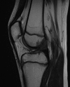
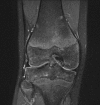
February is a busy month for the ambulance skiing patrol at the skiing resorts in Norway and on this day, a call regarding an 11-year-old boy on one of the hills reached the team. What no one knew at that moment was that this boy had suffered a unique injury and that his X-rays would reveal something that, prior to this, had never been described in the history of mankind. This patient had suffered a simultaneous avulsion fracture of both the femoral and tibial insertion sites of the anterior cruciate ligament without suffering any other injuries to the knee. The injury was treated conservatively and at 1-year follow-up, the patient was completely recovered.
Keywords: knee injuries; knee laxity; ligament rupture; orthopaedics; paediatrics.
© BMJ Publishing Group Ltd (unless otherwise stated in the text of the article) 2018. All rights reserved. No commercial use is permitted unless otherwise expressly granted.
Conflict of interest statement
Competing interests: None declared.
Figures


Similar articles
-
The Segond Fracture Is an Avulsion of the Anterolateral Complex.Am J Sports Med. 2017 Aug;45(10):2247-2252. doi: 10.1177/0363546517704845. Epub 2017 May 12. Am J Sports Med. 2017. PMID: 28499093
-
Cruciate ligament avulsion fractures.Arthroscopy. 2004 Oct;20(8):803-12. doi: 10.1016/j.arthro.2004.06.007. Arthroscopy. 2004. PMID: 15483540
-
Segond fracture.Joint Bone Spine. 2017 May;84(3):357. doi: 10.1016/j.jbspin.2016.06.008. Epub 2016 Jul 28. Joint Bone Spine. 2017. PMID: 27477317 No abstract available.
-
Femoral avulsion fracture of ACL proximal attachment in male scuba diver: case report and review of the literature.Knee Surg Sports Traumatol Arthrosc. 2017 Apr;25(4):1328-1330. doi: 10.1007/s00167-016-4373-x. Epub 2016 Nov 11. Knee Surg Sports Traumatol Arthrosc. 2017. PMID: 27837221 Review.
-
Knee and ankle injuries in children.Pediatr Rev. 1992 Nov;13(11):429-34. Pediatr Rev. 1992. PMID: 1289871 Review. No abstract available.
Cited by
-
ACL Repair of Femoral Osseous Avulsion in a 13-Year-Old Using Suture Pullout Technique.Video J Sports Med. 2021 Oct 5;1(5):26350254211030289. doi: 10.1177/26350254211030289. eCollection 2021 Sep-Oct. Video J Sports Med. 2021. PMID: 40308284 Free PMC article.
-
Transosseous suture in situ repair treatment of a femoral anterior cruciate ligament avulsion fracture in a 30-year-old male patient: a case report and review of the literature.Front Surg. 2025 Jul 10;12:1598881. doi: 10.3389/fsurg.2025.1598881. eCollection 2025. Front Surg. 2025. PMID: 40709060 Free PMC article.
-
A new fixation method for anterior cruciate ligament femoral avulsion fracture: a rare case report and literature review.Front Surg. 2025 Apr 8;12:1501740. doi: 10.3389/fsurg.2025.1501740. eCollection 2025. Front Surg. 2025. PMID: 40264741 Free PMC article.
References
Publication types
MeSH terms
LinkOut - more resources
Full Text Sources
Other Literature Sources
Medical
Miscellaneous
