. 2018 Mar-Apr;15(2):91-114.
doi: 10.21873/cgp.20068.

Characterization of Camptothecin-induced Genomic Changes in the Camptothecin-resistant T-ALL-derived Cell Line CPT-K5

Affiliations

Characterization of Camptothecin-induced Genomic Changes in the Camptothecin-resistant T-ALL-derived Cell Line CPT-K5

Eigil Kjeldsen et al. Cancer Genomics Proteomics. 2018 Mar-Apr.

Abstract

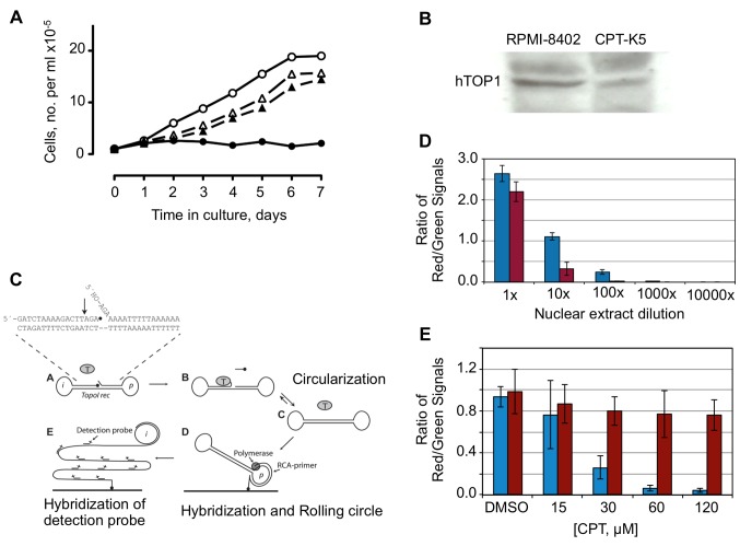
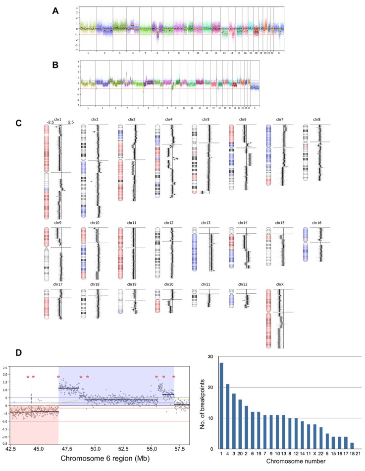
Acquisition of resistance to topoisomerase I (TOP1)-targeting camptothecin (CPT) derivatives is a major clinical problem. Little is known about the underlying chromosomal and genomic mechanisms. We characterized the CPT-K5 cell line expressing mutant CPT-resistant TOP1 and its parental T-cell derived acute lymphoblastic leukemia CPT-sensitive RPMI-8402 cell line by karyotyping and molecular genetic methods, including subtractive oligo-based array comparative genomic hybridization (soaCGH) analysis. Karyotyping revealed that CPT-K5 cells had acquired additional structural aberrations and a reduced modal chromosomal number compared to RPMI-8402. soaCGH analysis identified vast copy number alterations and >200 unbalanced DNA breakpoints distributed unevenly across the chromosomal complement in CPT-K5. In addition, the short tandem repeat alleles were found to be highly different between CPT-K5 and its parental cell line. We identified copy number alterations affecting genes important for maintaining genome integrity and reducing CPT-induced DNA damage. We show for the first time that short tandem repeats are targets for TOP1 cleavage, that can be differentially stimulated by CPT.

Keywords: CPT-K5; aCGH; camptothecin resistance; genomic instability; karyotype; short tandem repeat.


Figures

Figure 1
Figure 1. Fluorescence in situ hybridization (FISH) analysis of RPMI-8402 cell line using the SIL-TAL1 split-apart probe set showing a nucleus with 2F2G pattern indicating deletion of the SCL/TAL1-Interrupring locus (STIL) gene in two alleles and a normal fusion in the other two alleles. FISH analysis using of RPMI-8402 cell line using the LMO1-TCRD single fusion probe showing a nucleus with 2F2R2G pattern indicating fusion of the LIM domain only 1 (LMO1) and the Tcrd T cell receptor delta chain (TCRD) genes in two alleles and no fusion in the other two alleles. FISH analysis of CPT-K5 cell line using the TAL1-STIL split-apart probe showing a nucleus with 2F1G pattern indicating deletion of the STIL gene in one allele and a normal fusion in the other two alleles. FISH analysis using of CPT-K5 cell line using the LMO1-TCRD single fusion probe showing a nucleus with 3R3G pattern indicating no fusion of the genes.
Figure 2
Figure 2. A: Effects of camptothecin (CPT) on cell growth of parental RPMI-8402 and CPT-K5 cells. Growth of parental cells in the absence (l)and presence of 1 μM CPT (l). Growth of CPT-K5 in the absence (s) and presence of 1 μM CPT (s). B: Western blot of nuclear extracts from106 cells of RPMI-8402 and CPT-K5 using mouse antibody against human topoisomerase 1 (hTOP1). C: Schematic representation of the rollingcircle enhanced enzyme activity detection (REEAD) assay. A dumbbell-shaped DNA substrate with a preferred TOP1 cleavage sequence in the double-stranded stem region and an identifier- or primer annealing sequence (denoted “i” or “p”) in each of the loops is converted to a closed circle upon cleavage and ligation by TOP1. The generated circle is then hybridized to a surface anchored primer with a sequence matching the “p” region and subjected to rolling circle amplification by added phi-polymerase. The resulting tandem repeat rolling circle product is visualized at the single molecule level by hybridization of fluorescent labeled detection probes matching the “i” region. D: REEAD measurement of hTOP1 in nuclear extracts generated from 106 cells and assayed either undiluted (1x) or 10, 100, 1000, or 10,000 times diluted as indicated. E: The CPT sensitivity of hTOP1 in extracts from RPMI-8402 (blue bars) and CPT-K5 (red bars) was analyzed by measuring the activity using the REEAD assay in the presence of increasing concentrations of CPT as indicated. The bar chart shows the mean values from three independent experiments and the error bars indicate standard deviations.
Figure 3
Figure 3. G-Banding (A, C) and spectral karyotyping (B,D) of the RPMI-8402 (A,B) and CPT-K5 (C,D) cell lines showing representative karyotypes.
Figure 4
Figure 4. Genomic profiling using 180k oligo-based array comparative genomic hybridization (aCGH) analysis. A: Whole-genome view of RPMI- 8402 against normal female DNA pool. B: Whole-genome view of CPT-K5 against normal female DNA pool. C: Subtractive aCGH analysis where RPMI-8402 served as reference. To the right of the individual ideograms, microarray profiles of copy number gains and losses are depicted. Gain is indicated by blue and loss is indicated by red as an overlay on the ideogram. The log2 ratios for each chromosome are −2.5, 0, and +2.5 as illustrated for chromosome 1. D: Subtractive aCGH-based breakpoint analysis. Left panel: Magnified view of copy number profile of a region on chromosome 6 (pos. 42,500,000-58,000,000 bp) in which asterisks indicate determined putative DNA breakpoints. Focusing on the called regions of loss (red) and gain (blue) indicates two breakpoints, while taking copy number changes within the called regions into account, eight putative DNA break points can be determined. Right panel. Diagram showing the number of putative DNA break points determined on each chromosome.
Figure 5
Figure 5. Fluorescence in situ hybridization (FISH) validation of copy number changes at specific genomic regions important for the camptothecin resistance in CPT-K5 cells. Left hand-side panel: Genomic profiles of chromosome 20 (upper panel), chromosome 4 (middle panel) and chromosome 14 (lower panel) together with their respective ideograms beneath. Magnified genomic profile views of corresponding altered chromosomal regions are given below chromosome 20, chromosome 4, and chromosome 14 indicating deleted region at 20q12 containing topoisomerase 1 (TOP1) gene, highly amplified region at 4q22.1 containing ATP-binding cassette sub-family G member 2 (ABCG2) gene and amplified region at 14q31.3q32.11 containing the tyrosyl-DNA phosphodiesterase 1 (TDP1) gene, respectively. Blue and red bars around the ideograms indicate regions of gains and losses, respectively. Regions with high gains and losses are indicated by double bars in their respective color. A yellow bar in the copy number profile of chromosome 14 indicates the region of gain in the CR1-CR2/MMRU cell lines (see Table III). Right hand-side panel: FISH analyses confirm the genomic array comparative genomic hybridization (aCGH) findings in the RPMI-8402 and CPT-K5 cell lines on interphase nuclei with hybridization signals using the following probe sets as indicated by red and green marks on the respective ideograms in the left hand-side panel: i) Bacterial artificial chromosome (BAC)-based probe containing the TOP1 gene (red) and centromere 20 probe (green); ii) BAC-based probe RP11-368G2 (green) containing the ABCG2 gene together with centromere 4 probe (red) – to the right metaphase spreads represented by partial karyograms of chromosomes 4 and 12; and iii) BAC-based probe RP11-213O13 (red) containing the TDP1 gene together with subtelomeric 14-qter probe (green) - to the right metaphase spreads represented by partial karyograms of derivative chromosomes 11 (der(11)t(11;14)) and 15 (der(15)t(14;15)) for RPMI-8402 and partial karyograms of derivative chromosomes 5 (der(5)t(5;14)(q35;q32)) and 14 for CPT-K5.
Figure 6
Figure 6. Schematic overview of mechanisms implicated in repair of camptothecin (CPT)-induced DNA damage. BER: Base excision repair; BIR: breakage-induced replication; CPT: camptothecin or its derivatives; DSB: double-strand break; HR: homologous recombination; MMBIR: microhomology-mediated BIR; MRN: meiotic recombination 11 (MRE11)/DNA repair protein RAD50(RAD50)/nibrin (NBS1) complex; NER: nucleotide excision repair; NHEJ: non-homologous end-joining; NHR: non-homologous recombination; PTEN(−) indicates phosphatase and tensin homolog deficiency; SSB: single-strand DNA break. Open red circle indicates fork collision and repair mechanisms; Y-P: 3’-phosphotyrosyl.
Figure 7
Figure 7. Bar chart showing the mean of the results obtained when measuring relative tyrosyl-DNA phosphodiesterase 1 (TDP1) activity in whole-cell extracts from 1.0×10⁶ and 0.5×10⁶ RPMI-8402 (black bars) and CPT-K5 (grey bars) cells, as indicated in the figure, in three independent repetitions. Relative TDP1 activity in CPT-K5 cells was increased by a factor of 1.42 (p<0.001; unpaired t-test). The error bars indicate standard deviation.
Figure 8
Figure 8. Topoisomerase 1 (TOP1) DNA cleavage assay using short tandem repeat (STR) sequences as DNA substrate. The top panel shows the construction of the double-stranded oligonucleotide substrates. The grey box indicate the double-stranded STR alleles representing no match (None) D7S820, a partial match (m) D5S818, and a complete match (M) for thyroxin peroxidase (TPOX) as observed in the STR profiling of DNA from RPMI-8402 and CPT-K5 cell lines. These oligonucleotides were constructed so that the sequence of each of the three selected RPMI-8402 STR alleles were flanked by a common sequence identical in the three different substrates. Each construct was 5’-radiolabeled and subjected to human topoisomerase 1 (hTOP1) DNA cleavage with purified recombinant hTOP1 in the absence and presence of CPT before the products were separated by 12% polyacrylamide gel electrophoresis. Lanes 1, 4 and 7: incubation without enzyme and CPT; lanes 2, 5 and 8: incubation with hTOP1 in the absence of CPT; lanes 3, 6 and 9: incubation with hTOP1 and 10 μM CPT. Uncleaved substrates are marked (S) and *denotes hTOP1 DNA cleavage sites.

Similar articles

Cited by

References

    1. Kantarjian HM, Beran M, Ellis A, Zwelling L, O’Brien S, Cazenave L, Koller C, Rios MB, Plunkett W, Keating MJ. Phase I study of Topotecan, a new topoisomerase I inhibitor, in patients with refractory or relapsed acute leukemia. Blood. 1993;81:1146–1151. - PubMed
    1. Smith DH, Adams JR, Johnston SR, Gordon A, Drummond MF, Bennett CL. A comparative economic analysis of pegylated liposomal doxorubicin versus topotecan in ovarian cancer in the USA and the UK. Ann Oncol. 2002;13:1590–1597. - PubMed
    1. Tsavaris N, Kosmas C, Skopelitis H, Papadoniou N, Polyzos A, Zografos G, Adoniou E, Gryniatsos J, Felekouras E, Zacharakis M, Sigala F, Bacoyiannis C, Papastratis G, Papalambros E. Sequential administration of 5-fluorouracil (5FU)/leucovorin (LV) followed by irinotecan (CPT-11) at relapse versus CPT-11 followed by 5-FU/LV in advanced colorectal carcinoma. A phase III randomized study. Chemotherapy. 2007;53:282–291. - PubMed
    1. Riemsma R, Simons JP, Bashir Z, Gooch CL, Kleijnen J. Systematic Review of topotecan (Hycamtin) in relapsed small cell lung cancer. BMC Cancer. 2010;10:436. - PMC - PubMed
    1. Douillard JY, Cunningham D, Roth AD, Navarro M, James RD, Karasek P, Jandik P, Iveson T, Carmichael J, Alakl M, Gruia G, Awad L, Rougier P. Irinotecan combined with fluorouracil compared with fluorouracil alone as first-line treatment for metastatic colorectal cancer: a multicentre randomised trial. Lancet. 2000;355:1041–1047. - PubMed

MeSH terms