. 2018 Mar 2;359(6379):1042-1046.
doi: 10.1126/science.aaq1739.

Accurate computational design of multipass transmembrane proteins

Affiliations

Accurate computational design of multipass transmembrane proteins

Peilong Lu et al. Science. .

Abstract

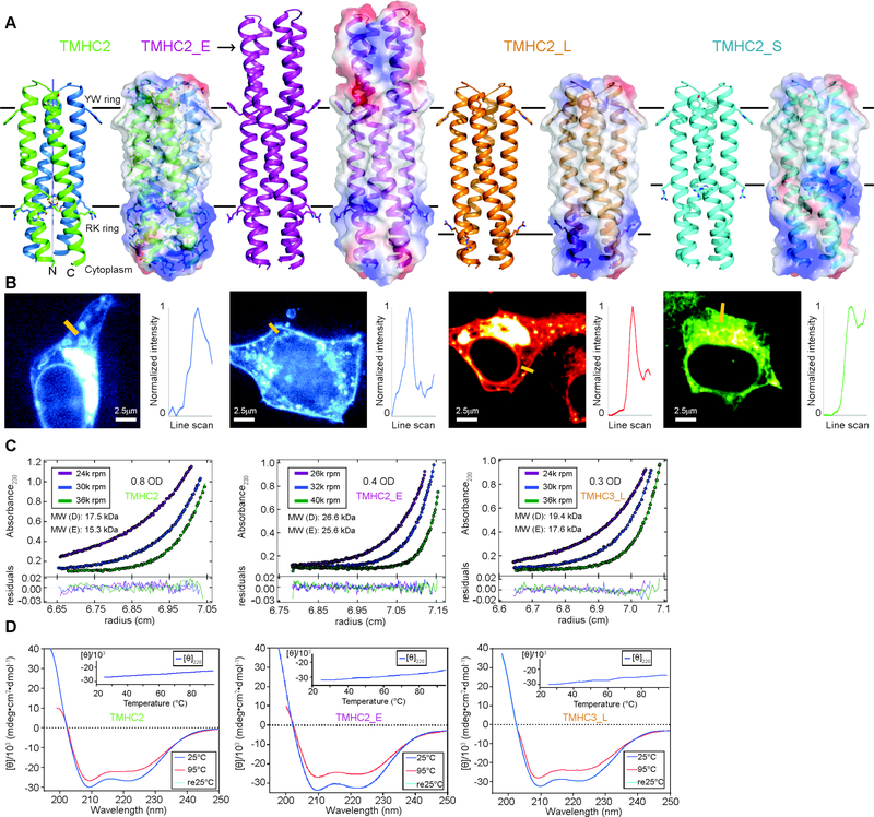
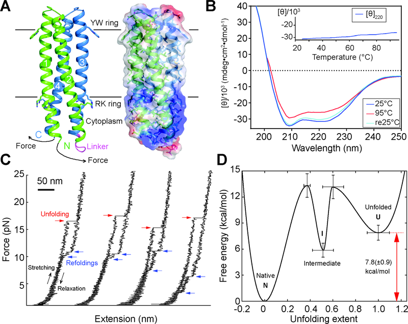
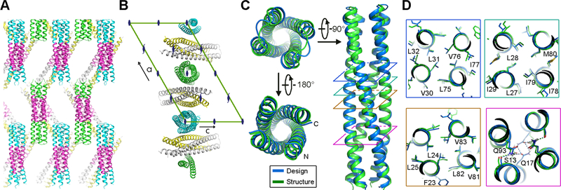
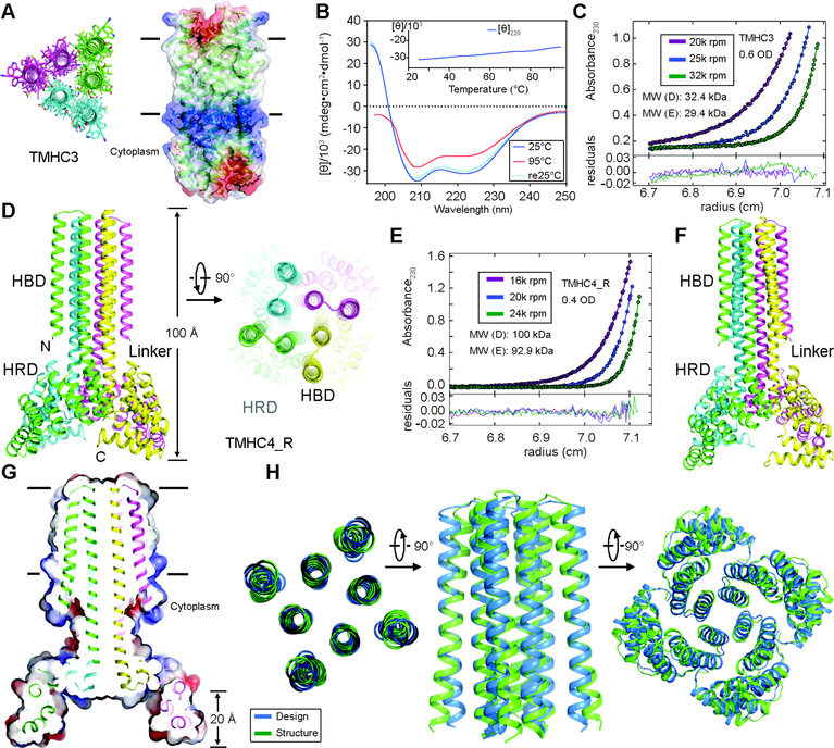
The computational design of transmembrane proteins with more than one membrane-spanning region remains a major challenge. We report the design of transmembrane monomers, homodimers, trimers, and tetramers with 76 to 215 residue subunits containing two to four membrane-spanning regions and up to 860 total residues that adopt the target oligomerization state in detergent solution. The designed proteins localize to the plasma membrane in bacteria and in mammalian cells, and magnetic tweezer unfolding experiments in the membrane indicate that they are very stable. Crystal structures of the designed dimer and tetramer-a rocket-shaped structure with a wide cytoplasmic base that funnels into eight transmembrane helices-are very close to the design models. Our results pave the way for the design of multispan membrane proteins with new functions.


Figures

Fig. 1.
Fig. 1.
Design and characterization of proteins with four transmembrane helices. From left to right, designs and data are shown for TMHC2 (transmembrane hairpin C2), TMHC2_E (elongated), TMHC2_L (long span) and TMHC2_S (short span). (A) Design models with intra- and extra-membrane regions with different lengths. Horizontal lines demarcate the hydrophobic membrane regions. Ribbon diagrams are on left, electrostatic surfaces on right, and the neutral transmembrane regions are in gray. (B) Confocal microscopy images for HEK293T cells transfected with TMHC2 fused to mTagBFP, TMHC2_E fused to mTagBFP, TMHC2_L fused to mCherry and TMHC2_S fused to eGFP. Line scans (yellow lines in the images) across the membranes show significant increase in fluorescence across the plasma membranes for TMHC2, TMHC2_E and TMHC2_L, but less significant increase for TMHC2_S. (C) Representative analytical ultracentrifugation sedimentation-equilibrium curves at three different rotor speeds. Each data set is globally well fitted as a single ideal species in solution corresponding to the dimer molecular weight. ‘MW (D)’ and ‘MW (E)’ indicate the molecular weight of the oligomer design and that determined from experiment, respectively. (D) CD spectra and temperature melt (inset). No apparent unfolding transitions are observed up to 95°C.
Fig. 2.
Fig. 2.
Folding stability of the 156-residue single chain TMHC2 (scTMHC2) design with four transmembrane helices. (A) Design model (left) and electrostatic surface (right) of scTMHC2. N- and C-terminal helical hairpins are colored green and blue respectively. Numbers indicate the order of the four TMs in the sequence. The linker connecting the two hairpins is colored magenta. Single-molecule forced unfolding experiments were conducted by applying mechanical tension to the N- and C-terminus of a single scTMHC2 (Fig. S5 for more details). (B) CD spectra of scTMHC2 at different temperatures. No unfolding transition is observed up to 95°C. (C) Single-molecule force-extension traces of scTMHC2. The unfolding and refolding transitions are denoted with red and blue arrows. (D) Folding energy landscape obtained from the single-molecule experiments. N, I, and U indicate the native, intermediate, and unfolded state respectively.
Fig. 3.
Fig. 3.
Crystal structure of the designed transmembrane dimer TMHC2_E. (A and B) Crystal lattice packing. (A) The extended soluble region mediates a large portion of the crystal lattice packing. The four helical hairpins in the asymmetric unit are colored green, gray, yellow and cyan, respectively. The TMs, in magenta, forms layers in the crystal separating the soluble regions. (B) The C2 axis of the design aligns with the crystallographic two fold. Two monomers (gray and yellow) are paired in a dimer while the other two (green and cyan) form two C2 dimers with two crystallographic adjacent monomers. The space group diagram (C121) is shown in the background. (C) Superposition of the TMHC2_E crystal structure and design model (RMSD = 0.7 Å over the core Cα atoms). (D) The side-chain packing arrangements at layers (colored squares in panel C) at different depths in the membrane are almost identical to the design model.
Fig. 4.
Fig. 4.
Stability and structural characterization of designs with six and eight membrane spanning helices. (A) Model of designed transmembrane trimer TMHC3 with six transmembrane helices. Stick representation from periplasmic side (left) and lateral surface view (right) are shown. (B) Circular dichroism characterization of TMHC3; the design is stable up to 95°C. (C) Representative analytical ultracentrifugation sedimentation-equilibrium curves at three different rotor speeds for TMHC3. The data fit to a single ideal species in solution with molecular weight close to that of the designed trimer. (D) Model of designed transmembrane tetramer TMHC4_R with eight transmembrane helices. The four protomers are colored green, yellow, magenta and cyan, respectively. (E) Analytical ultracentrifugation sedimentation-equilibrium curves at three different rotor speeds for TMHC4_R fit well to a single species with a measured molecular weight of ~94 kDa. (F) Crystal structure of TMHC4_R. The overall tetramer structures are very similar to the design model, with a helical bundle body and helical repeat fins. The outer helices of the transmembrane hairpins tilt off the axis by ~10°. (G) Cross section through the TMHC4_R crystal structure and electrostatic surface; the HRD forms a bowl at the base of the overall structure with a depth of ~20 Å. The transmembrane region is indicated in lines. (H) Three views of the backbone superposition of TMHC4_R crystal structure and design model.

References

    1. Huang P-S, Boyken SE, Baker D, The coming of age of de novo protein design. Nature. 537, 320–327 (2016). - PubMed
    1. Joh NH et al., De novo design of a transmembrane Zn2 -transporting four-helix bundle. Science. 346, 1520–1524 (2014). - PMC - PubMed
    1. Mahendran KR et al., A monodisperse transmembrane α-helical peptide barrel. Nat. Chem. 9, 411–419 (2016). - PubMed
    1. Whitley P, Nilsson I, von Heijne G, De novo design of integral membrane proteins. Nat. Struct. Mol. Biol. 1, 858–862 (1994). - PubMed
    1. Choma C, Gratkowski H, Lear JD, DeGrado WF, Asparagine-mediated self-association of a model transmembrane helix. Nat. Struct. Biol. 7, 161–166 (2000). - PubMed

Publication types