. 2018 Mar 1;9(3):338.
doi: 10.1038/s41419-018-0358-7.

Ischemic preconditioning attenuates ischemia/reperfusion-induced kidney injury by activating autophagy via the SGK1 signaling pathway

Affiliations

Ischemic preconditioning attenuates ischemia/reperfusion-induced kidney injury by activating autophagy via the SGK1 signaling pathway

Ying Xie et al. Cell Death Dis. .

Abstract

Ischemic preconditioning (IPC) has a strong renoprotective effect during renal ischemia/reperfusion (I/R) injury that is thought to relate to autophagy. However, the role of autophagy during IPC-afforded renoprotection and the precise mechanisms involved are unknown. In this study, an in vitro hypoxia/reoxygenation (H/R) model was established in which oxygen and glucose deprivation (OGD) was applied to renal cells for 15 h followed by reoxygenation under normal conditions for 30 min, 2 h or 6 h; transient OGD and subsequent reoxygenation were implemented before prolonged H/R injury to achieve hypoxic preconditioning (HPC). 3-Methyladenine (3-MA) was used to inhibit autophagy. In a renal I/R injury model, rats were subjected to 40 min of renal ischemia followed by 6 h, 12 h or 24 h of reperfusion. IPC was produced by four cycles of ischemia (8 min each) followed by 5 min of reperfusion prior to sustained ischemia. We found that IPC increased LC3II and Beclin-1 levels and decreased SQSTM/p62 and cleaved caspase-3 levels in a time-dependent manner during renal I/R injury, as well as increased the number of intracellular double-membrane vesicles in injured renal cells. IPC-induced renal protection was efficiently attenuated by pretreatment with 5 mM 3-MA. Pretreatment with IPC also dynamically affected the expression of SGK1/FOXO3a/HIF-1α signaling components. Moreover, knocking down SGK1 expression significantly downregulated phosphorylated-FOXO3a (p-FOXO3a)/FOXO3 and HIF-1α, suppressed LC3II and Beclin-1 levels, increased SQSTM/p62 and cleaved caspase-3 levels, and abolished the protective effect of IPC against I/R-induced renal damage. SGK1 overexpression efficiently increased p-FOXO3a/FOXO3 and HIF-1α levels, promoted the autophagy flux and enhanced the protective effect mediated by HPC. Furthermore, FOXO3a overexpression decreased HIF-1α protein levels, inhibited HIF-1α transcriptional activity and reduced the protective effect of IPC. Our study indicates that IPC can ameliorate renal I/R injury by promoting autophagy through the SGK1 pathway.


Conflict of interest statement

The authors declare that they have no conflict of interest.

Figures

Fig. 1
Fig. 1. Hypoxic preconditioning (HPC) protects renal tubular cells from hypoxia/reperfusion (H/R) injury by inducing autophagy in vitro.
a HK-2 cells were cultured under normal or oxygen and glucose deprivation (OGD) conditions for 15 h, followed by reoxygenation in normal medium for 2 h with or without prior HPC, which consisted of OGD for 6 h and subsequent reoxygenation for 2 h. The inhibitor 3-methyladenine (3-MA; 5 mM) was added to the medium 1 h before each experiment to suppress autophagy. Western blotting and subsequent data analysis were performed to ascertain LC3II, Beclin-1, SQSTM1/p62, and cleaved caspase-3 protein levels in HK-2 cells. Representative data are presented. b Determination and quantitative analysis of apoptotic cells by Annexin V-propidium iodide FACS analysis. c, d Renal tubular cell viability and lactic dehydrogenase (LDH) release were evaluated by CCK-8 and LDH assays. Each experiment was conducted at least three times independently. The data are presented as the mean ± S.D. n = 3*.P < 0.05
Fig. 2
Fig. 2. The SGK1/FOXO3a/HIF-1α signaling pathway is involved in HPC-mediated renal protection during H/R injury in vitro.
a HK-2 cells were cultured under normal or oxygen and glucose deprivation (OGD) conditions for 15 h, followed by reoxygenation in normal medium for 30 min, 2 h or 6 h with or without prior HPC, which consisted of OGD for 6 h and subsequent reoxygenation for 2 h. p-SGK1 and SGK1 protein levels were evaluated by western blotting. b, c Western blotting analysis of SGK1 knockdown and overexpression efficacy. Cells were transfected with Scramble, shRNA-SGK1 or pEX-3-SGK1. After 48 h, total protein was extracted for western blotting analysis. d, e Cells were transfected with Scramble, shRNA-SGK1 or pEX-3-SGK1. After 48 h, they were cultured under normal conditions or exposed to H/R injury with or without prior HPC. After reoxygenation for 2 h, cells were collected for western blotting detection of p-FOXO3a, FOXO3a and HIF-1α. Representative data are presented. f, g Renal tubular cell viability was evaluated by CCK-8 assay. h, i Flow cytometry was used to analyze the percentage of apoptotic cells. All the data are derived from an experiment that was repeated at least three times. The data are presented as the mean ± S.D. n = 3*.P < 0.05 vs. Control; #P < 0.05 vs. H/R group at the corresponding reoxygenation time; ##P < 0.05 vs. Scramble; **P < 0.05 vs. Scramble + H/R; &P < 0.05 vs. Scramble + HPC + H/R; +P < 0.05 vs. shRNA-SGK1 + H/R or SGK1-OE + H/R
Fig. 3
Fig. 3. The level of FOXO3a affects the expression of HIF-1α protein and HPC-conferred renoprotection during H/R injury in vitro.
a Cells were transfected with pEX-3-FOXO3a. After 48 h, they were cultured under normal conditions or exposed to H/R injury with or without prior HPC. After reoxygenation for 2 h, cells were collected for western blotting detection of HIF-1α. b EPO, HO-1 and Bnip3 mRNA levels were measured by real-time qPCR. c Renal tubular cell viability was evaluated by CCK-8 assay. d Flow cytometry was used to analyze the percentage of apoptotic cells. All the data are derived from an experiment that was repeated at least three times. The data are presented as the mean ± S.D. n = 3*.P < 0.05 vs. Scramble + H/R; &P < 0.05 vs. Scramble + HPC + H/R; +P < 0.05 vs. FOXO3a-OE + H/R
Fig. 4
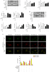
Fig. 4. HPC-mediated autophagy induction during renal H/R in vitro depends on SGK1.
a, b Cells were transfected with Scramble, shRNA-SGK1 or pEX-3-SGK1. After 48 h, they were cultured under normal conditions or exposed to H/R injury with or without prior HPC. After reoxygenation for 2 h, cells were collected for western blotting detection of LC3II, Beclin-1, SQSTM/p62, and cleaved caspase-3. c HK-2 cells were firstly infected with Scramble, shRNA-SGK1 or pEX-3-SGK1, and then infected with adenovirus encoding mRFP-GFP-LC3. After 12 h cells were exposed to H/R injury with or without prior HPC. After reoxygenation for 2 h, the cells were collected and observed under a confocal microscope. The data are derived from an experiment that was repeated at least three times and are presented as the mean ± S.D. n = 3*.P < 0.05 vs. Scramble + H/R; #P < 0.05 vs. Scramble + HPC + H/R; **P < 0.05##,P < 0.05 vs. Scramble + HPC + H/R
Fig. 5
Fig. 5. Ischemic preconditioning (IPC) induces autophagy and has a protective effect in an renal ischemia/reperfusion (I/R) model in vivo.
a Male Sprague–Dawley (SD) rats were subjected to sham operation, single IPC, or 40-min ischemia followed by 2-h, 6-h, 12-h or 24-h reperfusion with or without prior IPC, which consisted of four cycles of 8 min of clamping the left renal artery separated by 5 min of reperfusion. Western blotting and subsequent data analysis were performed to detect LC3II, Beclin-1, SQSTM1/p62 and cleaved caspase-3 protein levels. Representative data are presented. b Intracellular ultrastructural features of autophagy were observed by transmission electron microscopy (TEM). N: nucleus; arrowhead: double-membraned autophagosome; arrow: lysosome. Magnification, ×20,000; scale bar = 1 μm. c Serum creatinine (Scr): renal function parameter. Representative images of renal histology and quantitative analysis of tubular injury and apoptosis are presented. Hematoxylin-eosin (H&E) staining (d) and terminal deoxynucleotidyl transferase-mediated nick-end labeling (TUNEL) staining (e) of kidney sections. Representative slides from each group are presented. Magnification, ×200. All the data are derived from an experiment that was repeated at least three times and are presented as the mean ± S.D. n = 5*.P < 0.05 vs. Sham; #P < 0.05 vs. I/R group at the corresponding reperfusion time; **P < 0.05 vs. I/R-24 h
Fig. 6
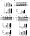
Fig. 6. Ischemic preconditioning (IPC) changes SGK1/FOXO3a/HIF-1α protein expression during renal I/R injury.
Male Sprague-Dawley (SD) rats were subjected to sham operation, single IPC, or 40-min ischemia followed by 2-h, 6-h, 12-h and 24-h reperfusion with or without prior IPC, which consisted of four cycles of 8 min of clamping the left renal artery separated by 5 min of reperfusion. a SGK1 mRNA levels in renal tissues from I/R-injured rats were measured by real-time qPCR and normalized to GAPDH expression. b Western blotting and subsequent data analysis were performed to detect p-SGK1 and SGK1 protein levels in renal tissues from I/R-injured rats. c, d Western blotting and data analysis were performed to detect p-SGK1, SGK1, p-FOXO3a, FOXO3a and HIF-1α expression levels in renal tissues from I/R-injured rats with or without prior IPC. Representative data are presented. All the data are derived from an experiment that was repeated at least three times and are presented as the mean ± S.D. n = 5 *.P < 0.05 vs. Sham; #P < 0.05 vs. I/R-6 h; ##P < 0.05 vs. I/R-24 h; **P < 0.05 vs. I/R group at the corresponding reperfusion time

Similar articles

Cited by

References

    1. Jiang M, et al. Autophagy in proximal tubules protects against acute kidney injury. Kidney Int. 2012;82:1271–1283. doi: 10.1038/ki.2012.261. - DOI - PMC - PubMed
    1. Kimura T, et al. Autophagy protects the proximal tubule from degeneration and acute ischemic injury. J. Am. Soc. Nephrol. 2011;22:902–913. doi: 10.1681/ASN.2010070705. - DOI - PMC - PubMed
    1. Joo J, Kim M, D’Agati V, Lee HT. Ischemic preconditioning provides both acute and delayed protection against renal ischemia and reperfusion injury in mice. J. Am. Soc. Nephrol. 2006;17:3115–3123. doi: 10.1681/ASN.2006050424. - DOI - PubMed
    1. Liu Z, Gong R. Remote ischemic preconditioning for kidney protection: GSK3β-centric insights into the mechanism of action. Am. J. Kidney Dis. 2015;66:846–856. doi: 10.1053/j.ajkd.2015.06.026. - DOI - PMC - PubMed
    1. El-Achkar T. Modulation of apoptosis by ischemic preconditioning: an emerging role for miR-21. Kidney Int. 2012;82:1149–1151. doi: 10.1038/ki.2012.305. - DOI - PubMed

Publication types

MeSH terms