. 2018 Mar 1;9(1):897.
doi: 10.1038/s41467-018-03355-0.

Conserved roles of C. elegans and human MANFs in sulfatide binding and cytoprotection

Affiliations

Conserved roles of C. elegans and human MANFs in sulfatide binding and cytoprotection

Meirong Bai et al. Nat Commun. .

Abstract

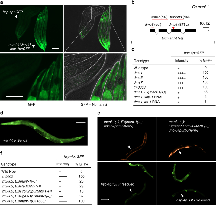
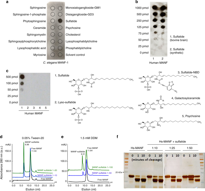
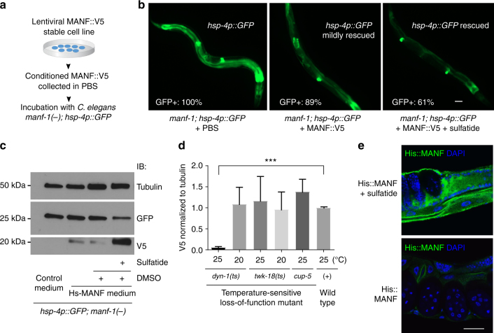
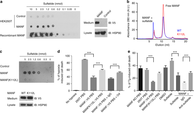
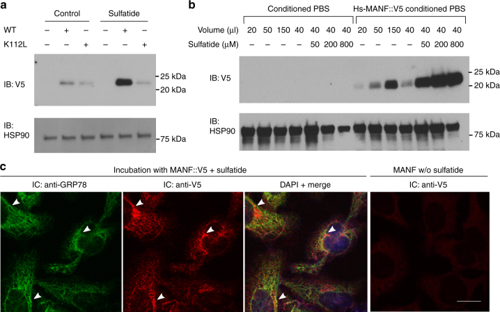
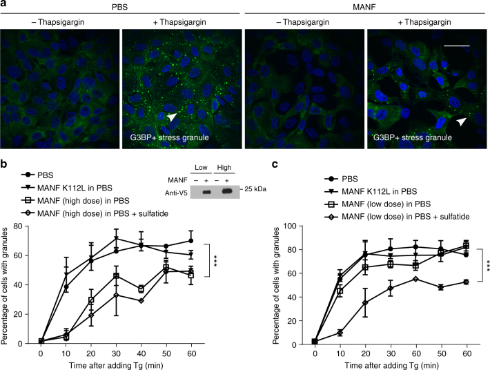
Mesencephalic astrocyte-derived neurotrophic factor (MANF) is an endoplasmic reticulum (ER) protein that can be secreted and protects dopamine neurons and cardiomyocytes from ER stress and apoptosis. The mechanism of action of extracellular MANF has long been elusive. From a genetic screen for mutants with abnormal ER stress response, we identified the gene Y54G2A.23 as the evolutionarily conserved C. elegans MANF orthologue. We find that MANF binds to the lipid sulfatide, also known as 3-O-sulfogalactosylceramide present in serum and outer-cell membrane leaflets, directly in isolated forms and in reconstituted lipid micelles. Sulfatide binding promotes cellular MANF uptake and cytoprotection from hypoxia-induced cell death. Heightened ER stress responses of MANF-null C. elegans mutants and mammalian cells are alleviated by human MANF in a sulfatide-dependent manner. Our results demonstrate conserved roles of MANF in sulfatide binding and ER stress response, supporting sulfatide as a long-sought lipid mediator of MANF's cytoprotection.


Conflict of interest statement

The authors declare no competing interests.

Figures

Fig. 1
Fig. 1
A genetic screen identifies C. elegans manf-1 in regulating hsp-4p::GFP. a Exemplar GFP fluorescence and Nomarski images showing constitutive activation of hsp-4p::GFP in the dma1 mutant isolated from mutagenesis screens. Wild type and mutant L4-stage animals (indicated by arrows) are aligned in the same field to compare GFP intensity at low (above) or high (bottom) magnification. Scale bars, 100 µM. b Schematic of manf-1 gene structure with various manf-1 alleles and extrachromosomal arrays (Ex) indicated. c Summary table for the hsp-4p::GFP phenotype of the wild type compared with various mutants, with indicated levels of GFP intensity in the intestine, the major site of hsp-4p::GFP expression, and penetrance (N ≥ 100 for each genotype). d Integrated manf-1p::Venus transcriptional reporter indicating the widespread expression of manf-1. e Exemplar GFP fluorescence images showing the rescue of the hsp-4p::GFP phenotype in the manf-1 null tm3603 mutant by C. elegans manf-1(+) and human MANF driven by the manf-1 promoter. Transgenic rescuing arrays are marked by unc-54p::mCherry expressed in body wall muscles. f Summary table showing the rescuing effect of various transgenes on hsp-4p::GFP of the manf-1(tm3603) mutant with indicated GFP intensity levels and penetrance (N ≥ 100 for each genotype)
Fig. 2
Fig. 2
Purified MANF binds to sulfatide. a Lipid overlay assay with an Echelon Sphingostrip membrane showing specific binding of His-tagged Ce-MANF to sulfatide. b Lipid overlay assay with customized lipid membrane showing dose-dependent binding of His-tagged Hs-MANF to sulfatide, prepared from bovine brains or synthetic source. c Lipid overlay assay with customized lipid membrane spotted with sulfatide-related lipids showing that sulfate as well as both acyl chains moieties are required for the sulfatide::Hs-MANF interaction. d Size exclusion chromatography with Hs-MANF reconstituted in 0.05% Tween-20 and varying doses of sulfatide. Molar ratio of MANF:Sulfatide at 1:20 was sufficient for 50% fraction to be bound onto tween-sulfatide micelles (peak at 6.99 ml elution). e Size exclusion chromatography with Hs-MANF reconstituted in 1.5 mM dodecyl maltoside (DDM) and varying doses of sulfatide. Molar ratio of MANF:Sulfatide at 1:100 resulted in 40% protein fraction to be bound onto DDM-sulfatide micelles (peak at 9.31 ml elution). f Limited proteolysis using trypsin protease showing that Hs-MANF is protected against digestion upon sulfatide binding, under conditions of different cleavage times (0, 1, and 10 min) and MANF versus sulfatide molar ratios (1:10, 1:25, and 1:50)
Fig. 3
Fig. 3
ER stress-rescuing effects of MANF and sulfatide in C. elegans. a Schematic of experimental workflow to test rescuing effects of human MANF from conditioned cell culture PBS. b Exemplar GFP fluorescence images showing the rescue of the hsp-4p::GFP phenotype in the manf-1 null tm3603 mutant by incubation with purified MANF secreted from conditioned media. MANF alone rescued only mildly compared with fuller rescue by MANF plus exogenous sulfatide addition (500 µM in DMSO; sulfatide alone had no rescue), with indicated penetrance of rescue (N ≥ 100 for each genotype). c Exemplar western blot of Hs-MANF in C. elegans manf-1 mutants after incubating with Hs-MANF, showing sulfatide dependency of endocytosis and rescue of hsp-4::GFP levels by endocytosed Hs-MANF. d Normalized levels of Hs-MANF::V5 immunoblot signals (from three biological replicates of N ≥ 100 animals) showing block of Hs-MANF endocytosis in temperature-sensitive endocytosis mutant dyn-1(ts) cultured from 25 °C but not at the permissive temperature, 20 °C. e Exemplar immunofluorescence images of C. elegans tissues stained with anti-His (green) and DAPI (blue) indicating the uptaken His::MANF with sulfatide. Scale bar, 15 µm
Fig. 4
Fig. 4
Sulfatide binding is critical for cytoprotection by MANF in culture. a Lipid overlay assay showing secreted V5-tagged Hs-MANF and recombinant His-tagged Hs-MANF bound to sulfatide (left), with secreted Hs-MANF::V5 confirmed by western blot of cell lysate and medium (right). HSP90 was used as a loading control. b Size exclusion chromatography of wild-type Hs-MANF and K112L mutants reconstituted in 0.05% Tween-20 and sulfatide. Only 20% of the K112L mutant Hs-MANF was bound onto the micelle fraction compared with 50% of the wild-type Hs-MANF. c Lipid overlay assay (above) showing that secreted MANF (K112L) from HEK293T cell lines exhibited markedly reduced binding to sulfatide with western blot (bottom) showing normal protein levels in the HEK293T stable line. d Death rates of HEK293T cells after 48 h of 0.1% hypoxia with pretreatment of Hs-MANF or MANF (K112L) mutant-conditioned media or with additional anti-sulfatide O4 antibodies. e Death rates of HEK293T cells after 24 h of indicated treatment followed by 40 h exposure to 0.5 µM thapsigargin (Tg). Error bars: S.E. with ***P < 0.001 (N ≥ 3 independent biological replicates)
Fig. 5
Fig. 5
Sulfatide promotes cellular uptake of MANF. a Exemplar western blot of lysates from target HEK293T cells treated with Hs-MANF or Hs-MANF (K112L) (purified from conditioned PBS of HEK293T-MANF::V5 cell lines) with or without sulfatide. b Exemplar western blot of lysates from target HEK293T cells treated with Hs-MANF (purified from conditioned PBS of HEK293T-MANF::V5 cell lines) with increasing doses of sulfatide added to the target HEK293T cell medium. c Exemplar immunofluorescence images of endogenous GPR78 (green) and V5-stained Hs-MANF (red), merged image with DAPI after incubation with Hs-MANF (purified from conditioned PBS of HEK293T-MANF::V5 cell lines) and with V5 staining of no-sulfatide control. Arrow heads indicate representative loci of co-localization. Scale bar, 20 µm
Fig. 6
Fig. 6
Sulfatide-bound MANF alleviates ER stress in mammalian cells. a Exemplar confocal GFP fluorescence images of Tg-induced stress granules in U2OS cells pretreated with conditioned MANF in PBS or control PBS. Scale bar, 40 µm. b Quantification of the percentages of cells with granules induced by Tg in 60 min and high doses (10 µl of conditioned medium/well) of Hs-MANF. A western blot for V5-tagged Hs-MANF indicates low and high doses of Hs-MANF used for experiments in b and c. c Quantification of the percentages of cells with granules induced by Tg in 60 min and low doses (2 µl of conditioned medium/well) of Hs-MANF. Cells were deprived of serum for 48 h and then treated with conditioned PBS or MANF for overnight. Plots represent quantification of the data from 10 images per condition from three independent experiments. Scale bar, 15 µm. *** indicates P < 0.001

References

    1. Tu BP, Weissman JS. Oxidative protein folding in eukaryotes: mechanisms and consequences. J. Cell Biol. 2004;164:341–346. doi: 10.1083/jcb.200311055. - DOI - PMC - PubMed
    1. Walter P, Ron D. The unfolded protein response: from stress pathway to homeostatic regulation. Science. 2011;334:1081–1086. doi: 10.1126/science.1209038. - DOI - PubMed
    1. Volmer R, Ron D. Lipid-dependent regulation of the unfolded protein response. Curr. Opin. Cell Biol. 2015;33:67–73. doi: 10.1016/j.ceb.2014.12.002. - DOI - PMC - PubMed
    1. Wouters BG, Koritzinsky M. Hypoxia signalling through mTOR and the unfolded protein response in cancer. Nat. Rev. Cancer. 2008;8:851–864. doi: 10.1038/nrc2501. - DOI - PubMed
    1. Semenza GL. Hypoxia-inducible factors in physiology and medicine. Cell. 2012;148:399–408. doi: 10.1016/j.cell.2012.01.021. - DOI - PMC - PubMed

Publication types