. 2018 Mar 1;8(1):3877.
doi: 10.1038/s41598-018-22093-3.

Landscape of the spliced leader trans-splicing mechanism in Schistosoma mansoni

Affiliations

Landscape of the spliced leader trans-splicing mechanism in Schistosoma mansoni

Mariana Boroni et al. Sci Rep. .

Abstract

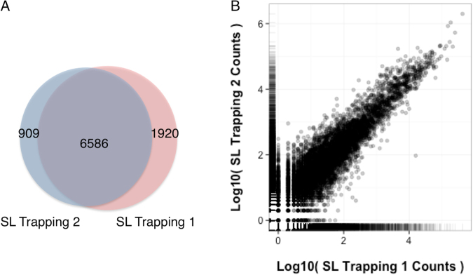
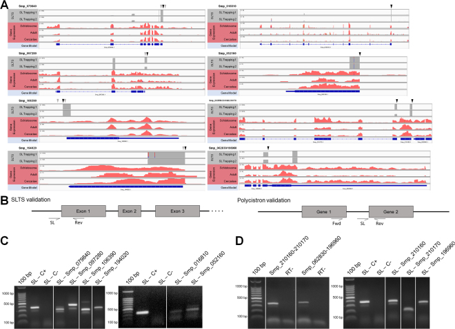
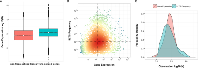
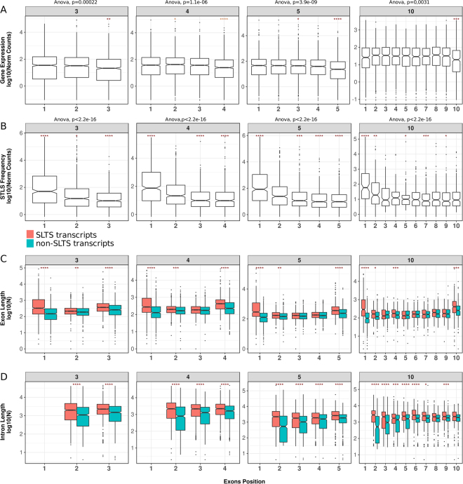
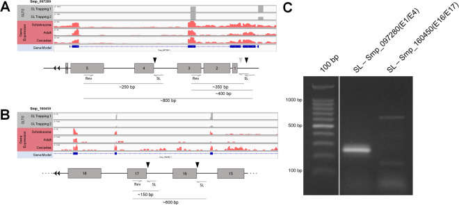
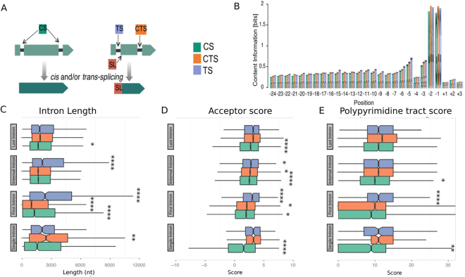
Spliced leader dependent trans-splicing (SLTS) has been described as an important RNA regulatory process that occurs in different organisms, including the trematode Schistosoma mansoni. We identified more than seven thousand putative SLTS sites in the parasite, comprising genes with a wide spectrum of functional classes, which underlines the SLTS as a ubiquitous mechanism in the parasite. Also, SLTS gene expression levels span several orders of magnitude, showing that SLTS frequency is not determined by the expression level of the target gene, but by the presence of particular gene features facilitating or hindering the trans-splicing mechanism. Our in-depth investigation of SLTS events demonstrates widespread alternative trans-splicing (ATS) acceptor sites occurring in different regions along the entire gene body, highlighting another important role of SLTS generating alternative RNA isoforms in the parasite, besides the polycistron resolution. Particularly for introns where SLTS directly competes for the same acceptor substrate with cis-splicing, we identified for the first time additional and important features that might determine the type of splicing. Our study substantially extends the current knowledge of RNA processing by SLTS in S. mansoni, and provide basis for future studies on the trans-splicing mechanism in other eukaryotes.


Conflict of interest statement

The authors declare no competing interests.

Figures

Figure 1
Figure 1
Comparison between the two SL Trapping experiments. (A) Shared genes between the two independent biological replicates, SL Trapping 1 (pink) and SL Trapping 2 (blue). (B) Correlation of gene counts between the two SL Trapping datasets (Pearson correlation coefficient = 0.86).
Figure 2
Figure 2
SL insertion sites and validation of novel trans-spliced transcripts. (A) Genome browser view of the exon-intron structure of genes undergoing SLTS, with the superimposed coverage for the SL Trapping 1 and 2 datasets (gray - top tracks) and the RNA-Seq dataset (pink - middle tracks). Black arrows show SL insertion sites and gray arrows show secondary SL insertions sites. The structure of genes with high (Smp_079840, Smp_097280, Smp_106390 and Smp_194020) and low read counts (Smp_016810 and Smp_052160), as well as the gene structure of a tricistronic transcript constituted by the genes Smp_033590, Smp_210160 and Smp_210170, and a dicistronic transcript, constituted by the genes Smp_062830 and Smp_196960, are shown. (B) Schematic view of genes validated by RT-PCR with the primers annealing sites. The expected fragment sizes were described in Table S1. (C) RT-PCR validation of trans-spliced genes with SL as forward primer and a gene-specific reverse primer. The Smp_024110 and Smp_045200 genes were included as positive and negative control, respectively, previously reported in. (D) RT-PCR validation of two polycistrons by two PCR reaction: with SL as the forward primer and a gene-specific reverse primer and with gene specific primers from upstream and downstream genes. Controls without reverse transcriptase were used for each of the reactions to show the absence of gDNA contamination. Full-length gels are presented in Supplementary Figure S5 A, B and C.
Figure 3
Figure 3
Relationship between gene expression and trans-splicing frequency. (A) Normalized log-expression levels of non-trans-spliced genes (pink box) compared with the log-expression levels of trans-spliced genes (blue box), p-value < 1.3e-08, two-sample t-test. (B) Normalized expression for the trans-spliced genes is compared with the normalized SLTS frequency. Dashed black lines denote the average levels of both indicators (163 for gene expression and 106 for SLTS frequency), segregating the genes with relatively low SLTS frequency (lower-right quadrant) from genes with relatively high SLTS frequency (upper-left quadrant) and comparing their expression levels (rank = 0.208, Spearman correlation coefficient). (C) The probability distribution of the gene expression levels (pink curve) and SLTS frequencies (blue curve) (p-value < 0.11, Kolmogorov-Smirnov).
Figure 4
Figure 4
Chromosomal origin, alternative splicing isoforms and number of exons in the trans-spliced genes. (A) The relative distribution of trans-spliced genes with high expression levels (GED, blue bars) and trans-spliced genes with low expression levels (TSD, purple bars) in each chromosome is compared with the chromosome distribution of genes expressed on the S. mansoni whole transcriptome (red bars) and genes expressed only in the cercariae transcriptome (green bars). (B) The relative distribution of alternative splicing transcript isoforms per gene in the S. mansoni whole transcriptome, in the cercariae transcriptome, in the subset of GED genes and in the subset of TSD genes. (C) The relative distribution of transcripts from genes with single or multiple exons in the S. mansoni whole transcriptome, in the cercariae transcriptome, in the subset of GED genes and in the subset of TSD genes.
Figure 5
Figure 5
Comparison between different positioning of acceptor sites in outrons and in introns subject to SLTS. (A) Relative number of cases and sequence logos of predicted acceptor sites in different positions according to the gene annotation. The positions of acceptor sites resulted from the mapping of SL Trapping reads. SLTS acceptor sites occurring in the first exon of transcripts, in the intron-exon junction, in the intron but upstream to the main acceptor site or located in the exon were named as outron, intron, alternative intron and exon, respectively. (B) Quantitative SLTS frequency in each of the different acceptor sites. ANOVA followed by pairwise t-test: ****p < = 0.0001. (C) Distribution of the dinucleotides found in each SLTS acceptor site positioning (pink bars - outron, green bars - intron, blue bars - alternative intron and purple bars - exon).
Figure 6
Figure 6
Trans-splicing occurrence by exons position in gene bodies and exon and intron lengths. (A) Expression levels of exons according to their position in transcripts (estimated from the RNA-Seq public dataset). (B) Frequency of trans-splicing according to exon position (estimated from SL Trapping). For A and B groups of genes were divided according to their number of annotated exons. Only genes with three, four, five and ten exons are shown. Analysis of variance (ANOVA) were performed followed by pairwise t-test (*p < = 0.05, **p < = 0.01,***p < = 0.001, ****p < = 0.0001). For a better visualization, pairwise differences were also tested by Tukey’s Honest significant difference test and the results are plotted on Figure S4. (C) Distribution of exon lengths in trans-spliced (pink boxes) and non-trans-spliced (blue boxes) genes that present three, four, five and ten exons. (D) Distribution of introns lengths immediately upstream of the splicing event in trans-spliced (pink boxes) and non-trans-spliced (blue boxes) genes presenting three, four, five and ten exons. For data in C and D, t-test were performed: *p < = 0.05, **p < = 0.01,***p < = 0.001, ****p < = 0.0001.
Figure 7
Figure 7
RT-PCR validation of internal trans-spliced genes. Genome browser and schematic view of the exon-intron structure of the genes Smp_062830 (A) and Smp_196960 (B) undergoing SLTS, with the superimposed coverage for the SL Trapping 1 and 2 datasets (top tracks) and the RNA-Seq dataset (middle tracks). Black arrows show the SL insertion sites and gray arrows show secondary SL insertion sites. The expected fragment sizes are described in the schematic representations. (C) RT-PCR validation of internal trans-splicing in the case studies using SL as forward primer and an exon-specific reverse primer. Full-length gels are presented in Supplementary Figure S5 D.
Figure 8
Figure 8
Cis and trans-splicing features based on the intron positions. (A) Introns were subdivided in three distinct groups for the following analysis: CS - cis-spliced introns (green boxes), CTS - cis-spliced introns in trans-spliced transcripts where the intron is not the primary SLTS target (orange boxes), and TS trans-spliced introns (purple boxes). (B) Intron/exons boundaries represented by the region from −24 to + 3 nucleotides. Acceptor splice sites in SL-containing genes were analysed based on their sequence composition. (C) Intron lengths are plotted based on their position in genes (single, first, internal, and last introns). (D) The strengths of acceptor splice sites (log-odds scores) are plotted according to the intron position in genes of the same three distinct groups. (E) Length of the polypyrimidine tract in nt relative to the acceptor sites are plotted according to intron position in genes of the same three distinct groups. Significantly differences are pointed (Kolmogorov-Smirnov test, *p < = 0.05, **p < = 0.01,***p < = 0.001, ****p < = 0.0001).

References

    1. Bitar M, Boroni M, Macedo AM, Machado CR, Franco GR. The spliced leader trans-splicing mechanism in different organisms: molecular details and possible biological roles. Front Genet. 2013;4:199. doi: 10.3389/fgene.2013.00199. - DOI - PMC - PubMed
    1. Thomas JD, Conrad RC, Blumenthal T. The C. elegans trans-spliced leader RNA is bound to Sm and has a trimethylguanosine cap. Cell. 1988;54:533–539. doi: 10.1016/0092-8674(88)90075-X. - DOI - PubMed
    1. Bruzik JP, Van Doren K, Hirsh D, Steitz JA. Trans splicing involves a novel form of small nuclear ribonucleoprotein particles. Nature. 1988;335:559–562. doi: 10.1038/335559a0. - DOI - PubMed
    1. Rajkovic A, Davis RE, Simonsen JN, Rottman FM. A spliced leader is present on a subset of mRNAs from the human parasite Schistosoma mansoni. Proc Natl Acad Sci USA. 1990;87:8879–8883. doi: 10.1073/pnas.87.22.8879. - DOI - PMC - PubMed
    1. Mourão M, et al. A directed approach for the identification of transcripts harbouring the spliced leader sequence and the effect of trans-splicing knockdown in Schistosoma mansoni. Mem Inst Oswaldo Cruz. 2013;108:707–717. doi: 10.1590/0074-0276108062013006. - DOI - PMC - PubMed

Publication types

LinkOut - more resources