. 2018 Mar 1;8(1):3863.
doi: 10.1038/s41598-018-22204-0.

Near-infrared laser thermal conjunctivoplasty

Affiliations

Near-infrared laser thermal conjunctivoplasty

Jianlong Yang et al. Sci Rep. .

Abstract

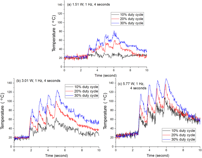
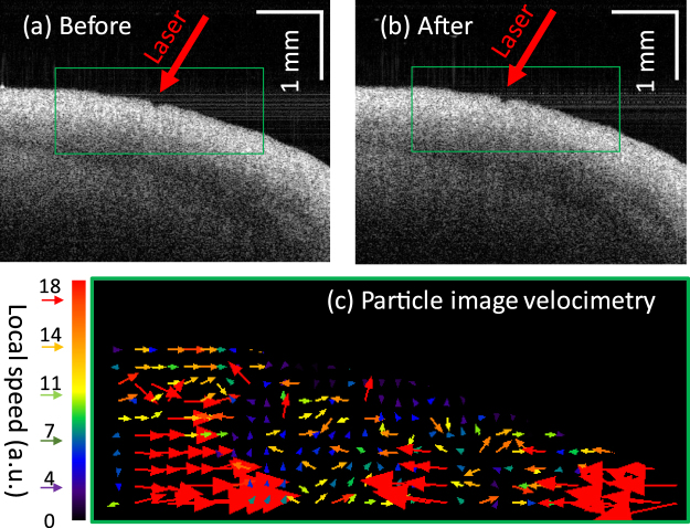
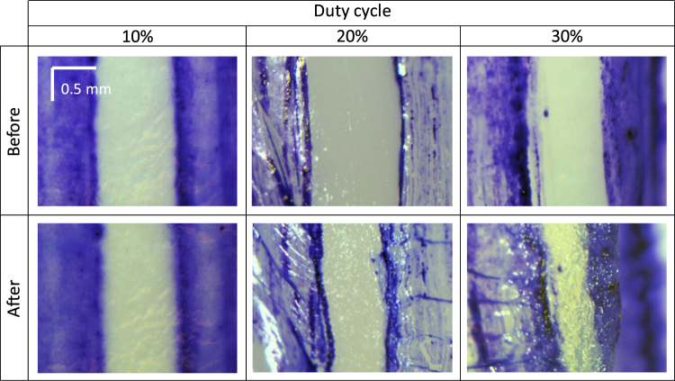
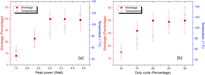
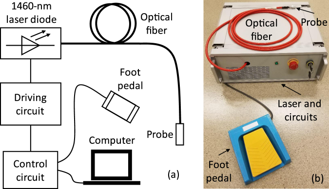
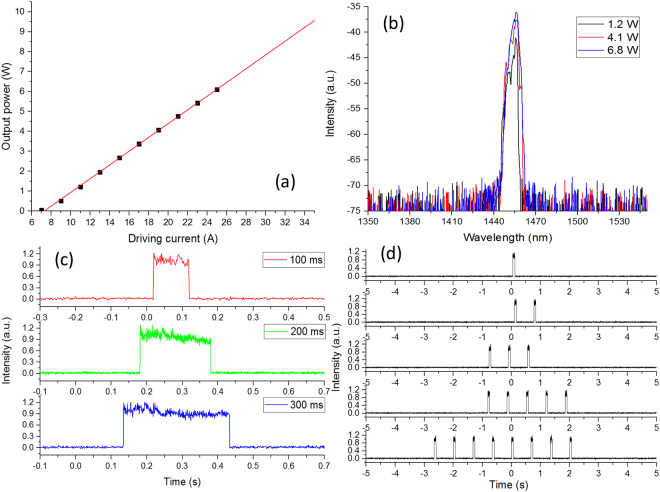
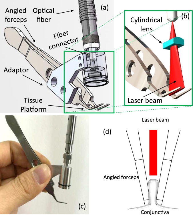
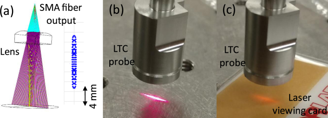
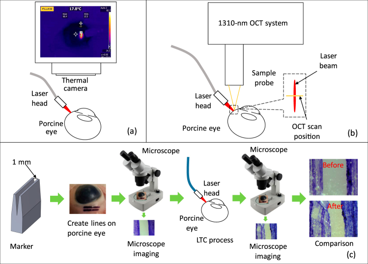
Conjunctivochalasis is a common cause of tear dysfunction due to the conjunctiva becoming loose and wrinkly with age. The current solutions to this disease include either surgical excision in the operating room, or thermoreduction of the loose tissue with hot wire in the clinic. We developed a near-infrared laser thermal conjunctivoplasty system. The system utilizes a 1460-nm programmable laser diode system as the light source. At this wavelength, a water absorption peak exists and the blood absorption is minimal, so the heating of redundant conjunctiva is even and there is no bleeding. A miniaturized handheld probe delivers the laser light and reshapes the laser into a 10 × 1 mm2 line on the working plane. A foot pedal is used to deliver a preset number of calibrated laser pulses. A fold of loose conjunctiva is grasped by a pair of forceps. The NIR laser light is delivered through an optical fiber and a laser line is aimed exactly on the conjunctival fold by a cylindrical lens. Ex vivo experiments using porcine eye was performed to investigate the induced shrinkage of conjunctiva and decide the optimal laser parameters. It was found that up to 45% of conjunctiva shrinkage could be achieved.


Conflict of interest statement

Oregon Health & Science University (OHSU), Baylor College of Medicine, David Huang, Gangjun Liu, Jianlong Yang, and Stephen C. Pflugfelder have potential financial interest in pending patent of the device described in the paper. These potential conflicts of interest have been reviewed and managed by OHSU.

Figures

Figure 1
Figure 1
Temperature changes during the LTC process using different laser parameters.
Figure 2
Figure 2
OCT B-frames taken before (a) and after (b) the LTC process (A video clip of the time-sequence OCT B-scans can be found in Supplementary information). (c) PIV images resulting from the green boxes in (a) and (b). The different colors correspond to different speeds and the arrows indicate the moving direction.
Figure 3
Figure 3
Photographs of the regions in the porcine eyes before and after LTC with different laser powers. The duty cycle is set at 20%. The blue regions are the marks from the tissue marker. The top row show the photographs before LTC and the corresponding photographs after the LTC are shown in the bottom row. Shrinkage of the tissue (the regions between the blue marks) could be seen from all the settings.
Figure 4
Figure 4
Photographs of the porcine eyes before and after LTC with different pulse duty cycle and a peak power of 3 W. The top row show the photographs before LTC and the corresponding photographs after the LTC are shown in the bottom row.
Figure 5
Figure 5
(a) Shrinkage and temperature as a function of laser peak power. The pulse laser duty cycle is set at 20%. The shrinkage rate saturates with the increase of the peak power and the temperature continues to increase with the increase of the peak power. (b) Shrinkage and temperature as a function of pulse duty cycle. The laser peak power was set as 3 W. The shrinkage rate saturates with the increase of the duty cycle and the temperature continues to increase with the increase of the duty cycle.
Figure 6
Figure 6
(a) Photograph of the LTC setup using the angled forceps. (b) The shrinkage results using only the forceps and both the forceps and the laser.
Figure 7
Figure 7
(a) Schematic of the 1460-nm LTC laser system. (b) Photograph of the prototype of the LTC laser system.
Figure 8
Figure 8
(a) Output power of the 1460-nm diode laser is proportional to the driving current. (b) The laser output spectrum at different output powers. (c) Temporal feature of output pulses with different pulse widths. (d) Output pulse trains with different pulse numbers.
Figure 9
Figure 9
(a) 3D model of the handheld probe with the angled forceps. (b) 3D demonstration of the line-focused laser beam and the angled forceps. (c) Photograph of the handheld LTC probe. (d) Illustration of the configuration for laser delivery to redundant conjunctival tissue.
Figure 10
Figure 10
(a) Simulation result of the laser beam transformation through the designed optics of the handheld LTC probe. (b) Photograph of the visible pilot laser from the handheld probe. (c) Photograph of the 1460-nm laser on a laser viewing card.
Figure 11
Figure 11
(a) Setup of measuring laser-induced temperature variation by a thermal camera. (b) Schematic of recording the dynamic shrinkage process by a 1310-nm optical coherence tomography system. (c) Procedure to measure the shrinkage from the LTC.

References

    1. Meller D, Tseng SCG. Conjunctivochalasis: Literature Review and Possible Pathophysiology. Survey of Ophthalmology. 1998;43(3):225–232. doi: 10.1016/S0039-6257(98)00037-X. - DOI - PubMed
    1. Liu D. Conjunctivochalasis: a cause of tearing and its management. Ophthalmic Plastic & Reconstructive Surgery. 1986;2(1):25–28. doi: 10.1097/00002341-198601040-00005. - DOI - PubMed
    1. Di Pascuale MA, Espana EM, Kawakita T, Tseng SCG. Clinical characteristics of conjunctivochalasis with or without aqueous tear deficiency. British journal of Ophthalmology. 2004;88:388–392. doi: 10.1136/bjo.2003.025460. - DOI - PMC - PubMed
    1. Zhang X, et al. Bulbar Conjunctival Thickness Measurements With Optical Coherence Tomography in Healthy Chinese Subjects. Investigative Ophthalmology & Visual Science. 2013;54:4705–4709. doi: 10.1167/iovs.12-11003. - DOI - PubMed
    1. Zhang Xingru, et al. In Vivo Cross-Sectional Observation and Thickness Measurement of Bulbar Conjunctiva Using Optical Coherence Tomography. Investigative Ophthalmology & Visual Science. 2011;52:7787–7791. doi: 10.1167/iovs.11-7749. - DOI - PubMed

Publication types