. 2018 Mar 1;8(1):3830.
doi: 10.1038/s41598-018-22301-0.

Acrylic Acid Plasma Coated 3D Scaffolds for Cartilage tissue engineering applications

Affiliations

Acrylic Acid Plasma Coated 3D Scaffolds for Cartilage tissue engineering applications

Pieter Cools et al. Sci Rep. .

Abstract

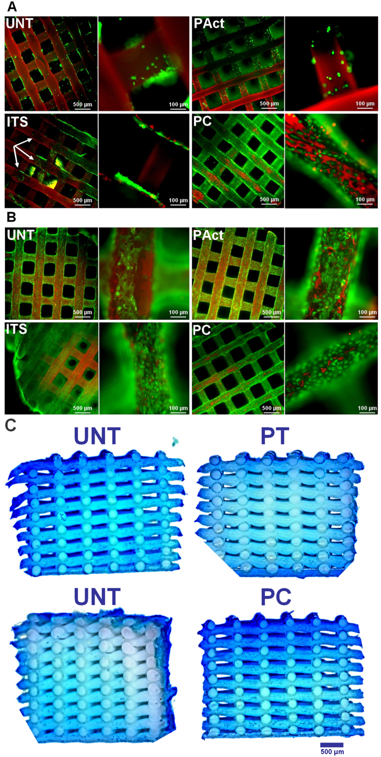
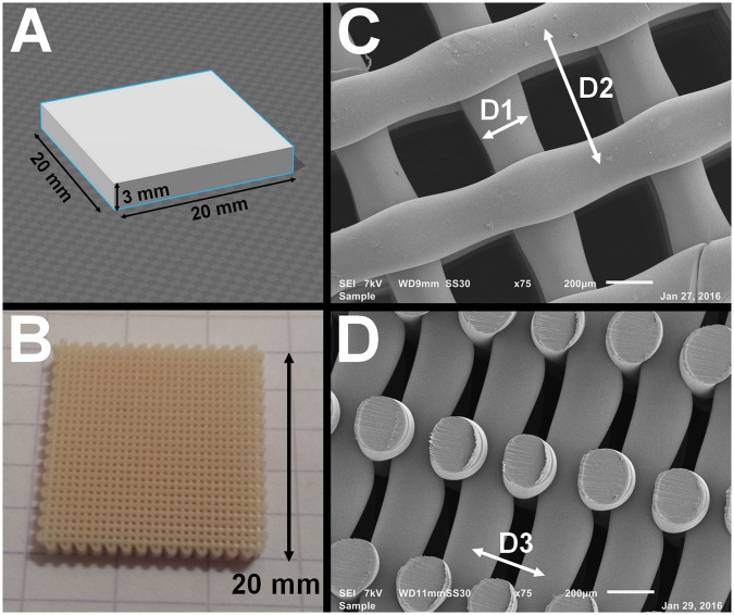
The current generation of tissue engineered additive manufactured scaffolds for cartilage repair shows high potential for growing adult cartilage tissue. This study proposes two surface modification strategies based on non-thermal plasma technology for the modification of poly(ethylene oxide terephthalate/poly(butylene terephthalate) additive manufactured scaffolds to enhance their cell-material interactions. The first, plasma activation in a helium discharge, introduced non-specific polar functionalities. In the second approach, a carboxylic acid plasma polymer coating, using acrylic acid as precursor, was deposited throughout the scaffolds. Both surface modifications were characterized by significant changes in wettability, linked to the incorporation of new oxygen-containing functional groups. Their capacity for chondrogenesis was studied using ATDC5 chondroblasts as a model cell-line. The results demonstrate that the carboxylic acid-rich plasma coating had a positive effect on the generation of the glucoaminoglycans (GAG) matrix and stimulated the migration of cells throughout the scaffold. He plasma activation stimulated the formation of GAGs but did not stimulate the migration of chondroblasts throughout the scaffolds. Both plasma treatments spurred chondrogenesis by favoring GAG deposition. This leads to the overall conclusion that acrylic acid based plasma coatings exhibit potential as a surface modification technique for cartilage tissue engineering applications.


Conflict of interest statement

The authors declare no competing interests.

Figures

Figure 1
Figure 1
(ac) XPS C1s deconvolution spectra for the untreated scaffold (a), the plasma activated scaffold (b) and the plasma coated scaffold (c). (AC) XPS measurements throughout the porous scaffold: (A) O1s concentration of the He plasma activated sample; (B) O1s concentration of the acrylic acid plasma coated sample; (C) O=C-O concentration as determined from deconvolution of the C1s peak (peak intensity at 289.1 eV) of the plasma coated sample.
Figure 2
Figure 2
Fluorescent micrographs of live/dead stained samples (4x and 20x) of the first set of experiments at time points day 1 (A) and day 15 (B) for the untreated samples (UNT), plasma activated samples (PAct), positive control (ITS) and plasma coated samples (PC). Methylene blue stained cross-sections of the scaffolds for the first set of experiments at day 15 (C).
Figure 3
Figure 3
Concentrations of DNA (A) and GAG (B) and their ratio (C) for the first set of experiments determined at fixed time points. *Significantly different compared to the negative control.
Figure 4
Figure 4
Methylene blue stained cross-sections of the scaffolds for the second set of experiments at day 1 (A); live/dead stained images for the second set of experiments at day 1 (B).
Figure 5
Figure 5
Concentrations of DNA (A) and GAG (B) and their ratio (C) for the second set of experiments determined at fixed time points. *Significantly different compared to the ITS containing sample.
Figure 6
Figure 6
SEM micrographs (top view (A) and cross-sectional view (B)) for the second set of experiments at day 5.
Figure 7
Figure 7
Fluorescent micrographs (left) and bright field (right) images (20x) of live/dead stained samples at time point day 10 (A); stereomicroscope images of methylene blue stained scaffolds (left: top view, right: cross-sectional view) at time point day 10 (B) and SEM micrographs (80x) (left: top view, right: cross-sectional view) of scaffolds at time point day 10 (C).
Figure 8
Figure 8
SEM micrographs of cross-sectioned scaffolds (80x and 200x) for the second set of experiments at time point day 15 (A) and methylene blue stained cross-sections of the second set of experiments at time point day 15 (B).
Figure 9
Figure 9
Collagen 2 concentration in the growth medium as determined via ELISA for the second set of experiments at time point day 15.
Figure 10
Figure 10
Visualization of the additive manufacturing process: (A) Model building in Google Sketch Up; (B) Photograph of the printed scaffold (1x magnification); (C) Top view SEM micrograph (75x magnification, 200 µm scale bar) showing the filament diameter D1 and the XY filament spacing D2; D) Cross-sectional SEM micrograph (75x magnification, 200 µm scale bar) showing the Z filament spacing.

References

    1. Lam J, et al. Evaluation of cell-laden polyelectrolyte hydrogels incorporating poly(l-Lysine) for applications in cartilage tissue engineering. Biomaterials. 2016;83:332–346. doi: 10.1016/j.biomaterials.2016.01.020. - DOI - PMC - PubMed
    1. Sophia Fox AJ, Bedi A, Rodeo SA. The Basic Science of Articular Cartilage: Structure, Composition, and Function. Sports Health: A Multidisciplinary Approach. 2009;1:461–468. doi: 10.1177/1941738109350438. - DOI - PMC - PubMed
    1. Raghunath J, Rollo J, Sales KM, Butler PE, Seifalian AM. Biomaterials and scaffold design: key to tissue-engineering cartilage. Biotechnol. Appl. Biochem. 2007;46:73–84. doi: 10.1042/BA20060134. - DOI - PubMed
    1. Camarero-Espinosa S, Rothen-Rutishauser B, Weder C, Foster EJ. Directed cell growth in multi-zonal scaffolds for cartilage tissue engineering. Biomaterials. 2016;74:42–52. doi: 10.1016/j.biomaterials.2015.09.033. - DOI - PubMed
    1. Zhang Y, et al. The impact of PLGA scaffold orientation on in vitro cartilage regeneration. Biomaterials. 2012;33:2926–2935. doi: 10.1016/j.biomaterials.2012.01.006. - DOI - PubMed

Publication types

MeSH terms

LinkOut - more resources