Review
. 2018 Feb 27;7(2):e1011.
doi: 10.1002/cti2.1011. eCollection 2018.

Unravelling the molecular basis for regulatory T-cell plasticity and loss of function in disease

Affiliations
Review

Unravelling the molecular basis for regulatory T-cell plasticity and loss of function in disease

Timothy Sadlon et al. Clin Transl Immunology. .

Abstract

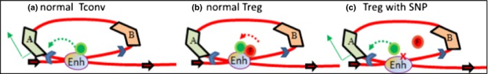
Regulatory T cells (Treg) are critical for preventing autoimmunity and curtailing responses of conventional effector T cells (Tconv). The reprogramming of T-cell fate and function to generate Treg requires switching on and off of key gene regulatory networks, which may be initiated by a subtle shift in expression levels of specific genes. This can be achieved by intermediary regulatory processes that include microRNA and long noncoding RNA-based regulation of gene expression. There are well-documented microRNA profiles in Treg and Tconv, and these can operate to either reinforce or reduce expression of a specific set of target genes, including FOXP3 itself. This type of feedforward/feedback regulatory loop is normally stable in the steady state, but can alter in response to local cues or genetic risk. This may go some way to explaining T-cell plasticity. In addition, in chronic inflammation or autoimmunity, altered Treg/Tconv function may be influenced by changes in enhancer-promoter interactions, which are highly cell type-specific. These interactions are impacted by genetic risk based on genome-wide association studies and may cause subtle alterations to the gene regulatory networks controlled by or controlling FOXP3 and its target genes. Recent insights into the 3D organisation of chromatin and the mapping of noncoding regulatory regions to the genes they control are shedding new light on the direct impact of genetic risk on T-cell function and susceptibility to inflammatory and autoimmune conditions.

Keywords: Treg FOXP3; T‐cell fate; T‐cell plasticity; gene regulation; genetic risk of disease; microRNA.


Figures

Figure 1
Figure 1
Functional annotation of FOXP3‐bound mRNA regions of the human genome intersected with differential mRNA expression (left Venn diagram), and FOXP3‐bound microRNA regions of the human genome intersected with differential miR expression (right Venn diagram), showing that a subset of mRNAs and miRs are both differentially expressed and FOXP3 targets.
Figure 2
Figure 2
(a) Schematic representation of the human FOXP3 gene, annotating the three conserved noncoding sequences involved in regulating gene expression (CNS1, 2, 3). (b) A VISTA alignment of mouse and human FOXP3 showing regulatory regions which are conserved between species in red, conserved untranslated regions in light blue and conserved exons in lilac.
Figure 3
Figure 3
FOXP3‐ and FOXP3‐induced microRNAs (green) cooperate to tightly repress (red) target genes in Treg, to reinforce the suppressor phenotype. These genes are actively expressed in effector T cells.
Figure 4
Figure 4
Molecular model of the FOXP3 gene regulatory network feedforward loops. FOXP3 represses key effector function genes both by direct binding to regulatory elements associated with the gene (e.g. SATB1) and by inducing miRs that themselves target the same gene 3′ UTR. In addition, FOXP3 induces Treg functional genes to reinforce the Treg lineage and phenotype, while suppressing inducers of effector lineage commitment.
Figure 5
Figure 5
Molecular model of the FOXP3 gene regulatory network feedback loop controlling FOXP3 expression. In a stable Treg, FOXP3 represses miR31 by direct binding to regulatory elements associated with the gene, and in stable effector T cells, miR31 expression prevents the expression of FOXP3 by targeting FOXP3 mRNA for degradation. Subtle alterations in levels of either miR31 or FOXP3 will have reciprocal effects on the expression of each, providing a threshold for transition between FOXP3 +ve and −ve states.
Figure 6
Figure 6
Integrated model of the plasticity of phenotypes based on molecular switching driven by induction of miRs which regulate the sentinel transcription factor for one phenotype and allow expression of the transcription factor driving the other phenotype. In regulatory T cells, a suite of FOXP3‐induced miRs are expressed in the steady state, but as a result of external cues such as cytokine signalling, these miRs are reduced, and competing miRs including miR31 are increased, reducing FOXP3 expression and releasing other transcription factors from repression, facilitating a transition and reversible functional switching.
Figure 7
Figure 7
Integrated model of regulation of effector function in effector T cells based on molecular switching driven by transient repression of miR(s) which de‐represses FOXP3, therefore transiently repressing effector function genes. This enables shut down of an immune response upon clearance of the pathogen.
Figure 8
Figure 8
Conformation‐dependent interactions contribute to gene regulation networks, and these are not readily predicted by bioinformatics approaches. (a) Linear annotation of TF binding site data to nearest neighbour is only accurate if within a short distance from the TSS. (b) unidentified FOXP3 binding sites, and distal regulatory interactions are annotated after incorporation of DNA looping data from conformation capture experiments.
Figure 9
Figure 9
Functional annotation of genetic risk from the GWAS data set which maps to FOXP3 ChIP binding regions. The addition of chromatin conformation data allows identification of the target genes, and this enables analysis of the impact of genetic variation. In a Tconv, the gene is expressed because there is no FOXP3 (a). In a normal Treg, the gene is repressed (b), but in a Treg from the disease cohort, the sequence variation results in loss of a FOXP3 binding site, as a result the target gene is no longer repressed (c). The functional impact is at a distal regulatory element, not in the target gene itself.

References

    1. Josefowicz SZ, Lu LF, Rudensky AY. Regulatory T cells: mechanisms of differentiation and function. Annu Rev Immunol 2012; 30: 531–564. - PMC - PubMed
    1. Shevach EM, Thornton AM. tTregs, pTregs, and iTregs: similarities and differences. Immunol Rev 2014; 259: 88–102. - PMC - PubMed
    1. Yadav M, Stephan S, Bluestone JA. Peripherally induced tregs ‐ role in immune homeostasis and autoimmunity. Front Immunol 2013; 4: 232. - PMC - PubMed
    1. Szymczak‐Workman AL, Workman CJ, Vignali DA. Cutting edge: regulatory T cells do not require stimulation through their TCR to suppress. J Immunol 2009; 182: 5188–5192. - PMC - PubMed
    1. Yamaguchi T, Wing JB, Sakaguchi S. Two modes of immune suppression by Foxp3(+) regulatory T cells under inflammatory or non‐inflammatory conditions. Semin Immunol 2011; 23: 424–430. - PubMed