. 2018 Feb 15:8:33.
doi: 10.3389/fonc.2018.00033. eCollection 2018.

Olfactory Receptors as Biomarkers in Human Breast Carcinoma Tissues

Affiliations

Olfactory Receptors as Biomarkers in Human Breast Carcinoma Tissues

Lea Weber et al. Front Oncol. .

Abstract

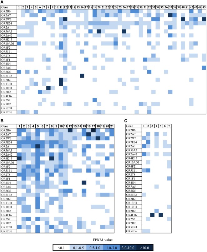
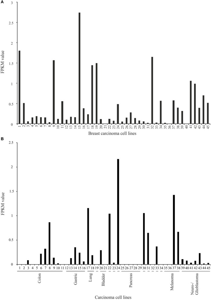
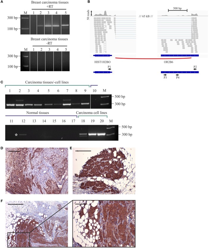
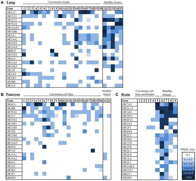
Olfactory receptors (ORs) are known to be expressed in a variety of human tissues and act on different physiological processes, such as cell migration, proliferation, or secretion and have been found to function as biomarkers for carcinoma tissues of prostate, lung, and small intestine. In this study, we analyzed the OR expression profiles of several different carcinoma tissues, with a focus on breast cancer. The expression of OR2B6 was detectable in breast carcinoma tissues; here, transcripts of OR2B6 were detected in 73% of all breast carcinoma cell lines and in over 80% of all of the breast carcinoma tissues analyzed. Interestingly, there was no expression of OR2B6 observed in healthy tissues. Immunohistochemical staining of OR2B6 in breast carcinoma tissues revealed a distinct staining pattern of carcinoma cells. Furthermore, we detected a fusion transcript containing part of the coding exon of OR2B6 as a part of a splice variant of the histone HIST1H2BO transcript. In addition, in cancer tissues and cell lines derived from lung, pancreas, and brain, OR expression patterns were compared to that of corresponding healthy tissues. The number of ORs detected in lung carcinoma tissues was significantly reduced in comparison to the surrounding healthy tissues. In pancreatic carcinoma tissues, OR4C6 was considerably more highly expressed in comparison to the respective healthy tissues. We detected OR2B6 as a potential biomarker for breast carcinoma tissues.

Keywords: OR2B6; RNA-Seq; biomarker; breast carcinoma; next-generation sequencing; olfactory receptors; pancreatic carcinoma.


Figures

Figure 1
Figure 1
Expression pattern of ectopically expressed olfactory receptors (ORs) in breast carcinoma tissue samples and cell lines and healthy breast tissue samples. (A) Heat map shows 26 ORs in 45 breast cancer cell lines. For further details on the cell lines, see Table S1 in Supplementary Material. Sample no. 1 and 45 are self-generated RNA-Seq data of breast cancer cell lines. (B) Heat map shows 26 ORs in 21 breast carcinoma tissues. Sample 1–13 are reanalyzed data sets from NCBI (for further details, see Section “Materials and Methods”), samples 15–21 are self-generated RNA-Seq data of breast carcinoma tissue samples. (C) Heat map shows 26 ORs in 7 normal breast tissue samples. Color intensity correlates with the expression strength. Dark blue represents FPKM values >3, and light blue represents FPKM values <0.5.
Figure 2
Figure 2
Expression analysis of OR2B6 in healthy tissue samples and cell lines. (A) Expression of OR2B6 in ten healthy cell lines originated from different human tissues. For further details see Table S2 in Supplementary Material. (B) Expression of OR2B6 in 16 different human tissues, origin: BodyMap Project (26). (C) Box-plot showing the expression of OR2B6 in normal breast tissues and breast carcinoma tissues and cell lines. Breast tissue: normal breast tissue samples, n = 7; breast tissue GTEx: normal breast tissue samples originated from GTEx database, n = 214; breast cell lines: cell lines originated from normal breast tissues, n = 4; breast carcinoma tissues: carcinoma breast tissue samples, n = 21; and breast carcinoma cell lines: cell lines originated from breast cancer tissues, n = 45. Normality test (Shapiro–Wilk) failed, Kruskal–Wallis one-way ANOVA: p < 0.05; the statistical significance is indicated as follows: **p < 0.01. Red line represents the median value. (D) Expression of OR2B6 in 52 healthy tissues originated from the GTEx database.
Figure 3
Figure 3
Expression analysis of OR2B6 in breast carcinoma cell lines and carcinoma cell lines that originated from other carcinoma types. (A) Expression of OR2B6 in 45 breast carcinoma cell lines. For further details on cell lines, see Table S1 in Supplementary Material. (B) Expression of OR2B6 in 45 other carcinoma cell lines, except breast carcinoma cell lines. 19: cell line that originated from osteosarcoma; 22: cell line that originated from prostate carcinoma; 23: cell line that originated from leukemia cells; 24: cell line that originated from germ cell carcinoma; 30: cell line that originated from cervix carcinoma; 31: cell line that originated from synovial sarcoma; 32: cell line that originated from Ewing sarcoma; 33: cell line that originated from myosarcoma; 34: cell line that originated from fibrosarcoma. For further details on cell lines, see Table S3 in Supplementary Material.
Figure 4
Figure 4
Expression of OR2B6 in human breast carcinoma tissues and detection of fusion transcripts by reverse transcriptase PCR (RT-PCR). (A) Validation of OR2B6 expression in five different breast carcinoma tissues via RT-PCR. +, +RT, cDNA; −, −RT, RNA; M, marker; bp, base pairs. (B) Visualization of the fusion transcript of OR2B6 and HIST1H2BO via the integrative genomic viewer. (C) Validation of the expression of the fusion transcript of OR2B6 and HIST1H2BO via RT-PCR. +, +RT, cDNA; −, −RT, RNA; M, marker; bp, base pairs. (D) Immunhistochemical staining of metaplastic breast carcinoma tissue using specific human α-OR2B6 antibody. Tumor details: G3, triple negative. Shown is the expression of the receptor protein in a triple-negative metaplastic breast carcinoma in G3. Cancer cells are specifically stained, surrounding connective tissue fibers and fat cells show no staining. (E) Immunhistochemical staining of invasive ductal breast carcinoma tissue using specific human α-OR2B6 antibody. Shown is the expression of the receptor in an E+/PR/HER2 carcinoma in G2 (estrogen 100%: ER+, progesterone 0%: PR, Her2 negative: HER2). Protein expression is visualized using DAB chromogenic staining and co-staining with hematoxylin (HE) is used to illustrate the tissue architecture. (F) Immunhistochemical staining of invasive ductal breast carcinoma tissue using specific human α-OR2B6 antibody. Tumor details: G3, triple negative. Shown is the expression of the receptor protein in a triple-negative invasive ductal breast carcinoma in G3. Scale bar: 100 µm.
Figure 5
Figure 5
Expression pattern of ectopically expressed olfactory receptors (ORs) in lung, pancreas, and brain carcinoma tissue samples, cell lines, and healthy tissues. (A) Heat map shows 20 ranked ORs according to their expression level in 20 lung carcinoma tissues in comparison to 5 healthy lung tissues. 1–20: lung carcinoma tissue samples; 21–25: normal lung tissue samples. (B) Heat map shows 20 ORs ranked according to their expression level in 20 pancreatic cancer tissues and cell lines in comparison to 2 healthy pancreas tissues. 1: Cell line Capan-1. 2–20: patient-derived pancreatic carcinoma cell lines. 21–22: healthy pancreas tissue samples. (C) Heat map shows 20 ORs ranked according to their expression level in 4 brain tumor tissue samples and cell lines and 5 healthy brain tissue samples. 1–3 brain tumor cell lines, 4: brain carcinoma tissue sample, 5–9: healthy brain tissue samples. Color intensity correlates with the expression strength. Dark blue represents FPKM values >3, and light blue represents FPKM values <0.5. For further details on cell lines, see Table S4 in Supplementary Material.

Comment in

References

    1. Firestein S. How the olfactory system makes sense of scents. Nature (2001) 413(6852):211–8.10.1038/35093026 - DOI - PubMed
    1. Glusman G, Yanai I, Rubin I, Lancet D. The complete human olfactory subgenome. Genome Res (2001) 11(5):685–702.10.1101/gr.171001 - DOI - PubMed
    1. Buck L, Axel R. A novel multigene family may encode odorant receptors: a molecular basis for odor recognition. Cell (1991) 65(1):175–87.10.1016/0092-8674(91)90418-X - DOI - PubMed
    1. Kang N, Koo J. Olfactory receptors in non-chemosensory tissues. BMB Rep (2012) 45(11):612–22.10.5483/BMBRep.2012.45.11.232 - DOI - PMC - PubMed
    1. Veitinger S, Hatt H. Ectopic Expression of Mammalian Olfactory Receptors. In: Buettner, Andrea editors. Springer Handbook of Odor, Springer Handbooks. Cham: Springer; (2017), p. 83–84.