. 2018 Feb 28;5(1):ENEURO.0240-17.2018.
doi: 10.1523/ENEURO.0240-17.2018. eCollection 2018 Jan-Feb.

α-Tubulin Acetyltransferase Is a Novel Target Mediating Neurite Growth Inhibitory Effects of Chondroitin Sulfate Proteoglycans and Myelin-Associated Glycoprotein

Affiliations

α-Tubulin Acetyltransferase Is a Novel Target Mediating Neurite Growth Inhibitory Effects of Chondroitin Sulfate Proteoglycans and Myelin-Associated Glycoprotein

Victor S C Wong et al. eNeuro. .

Abstract

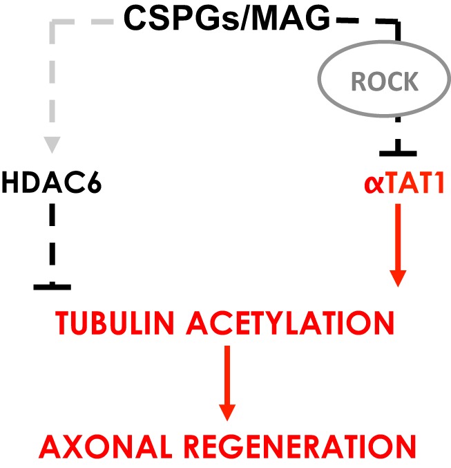
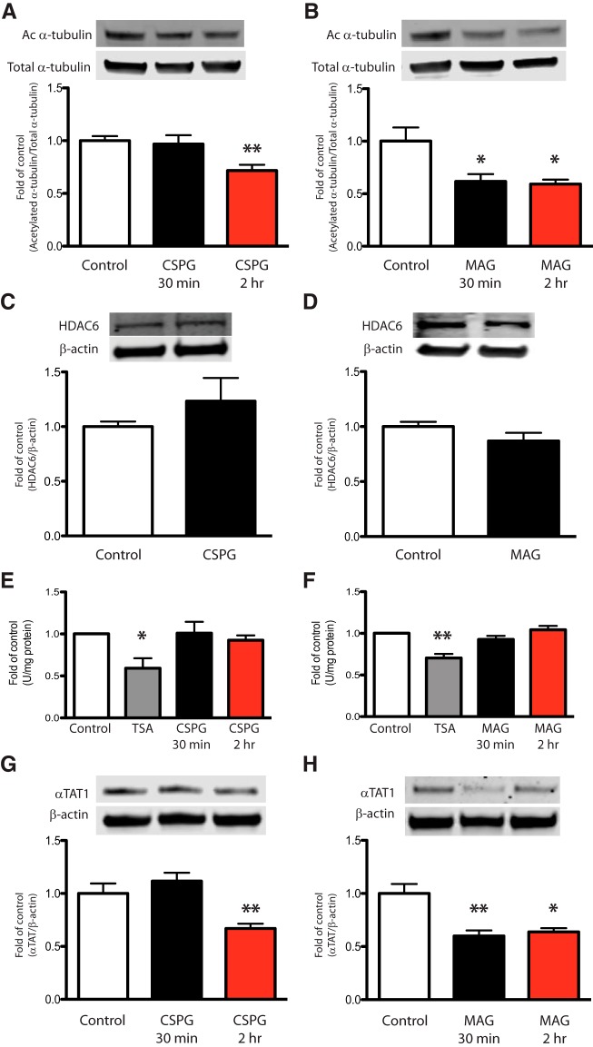
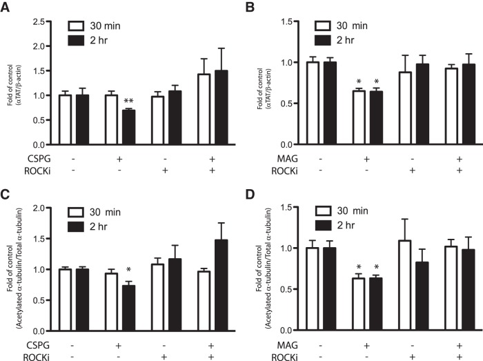
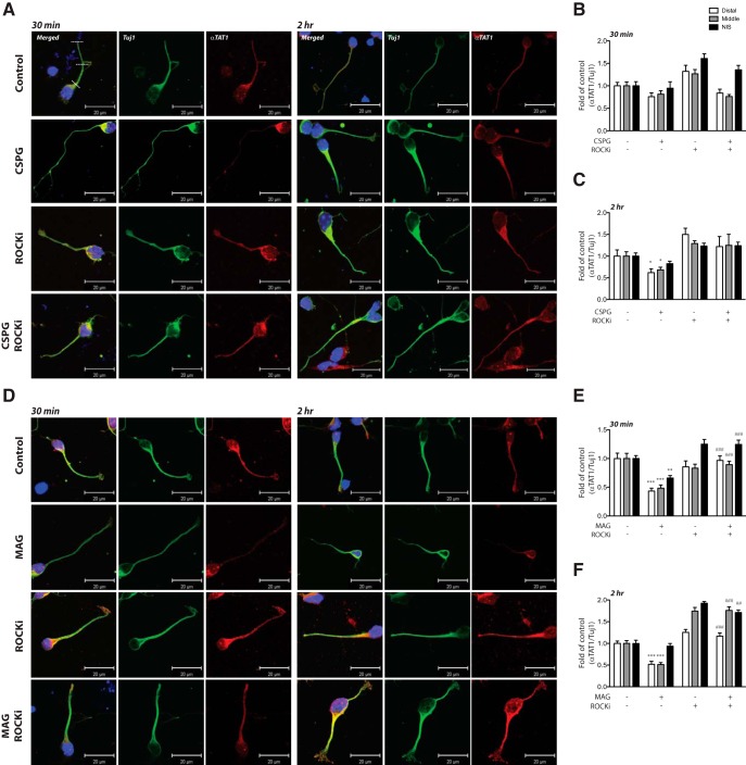
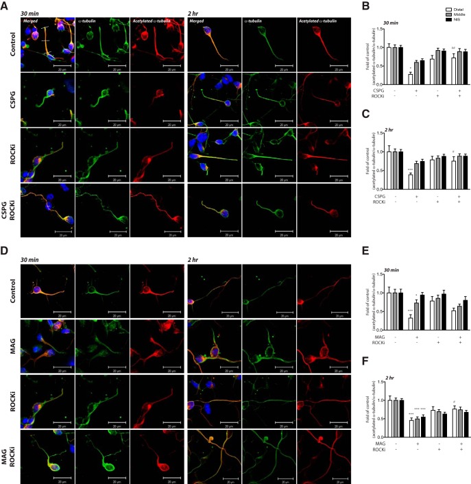
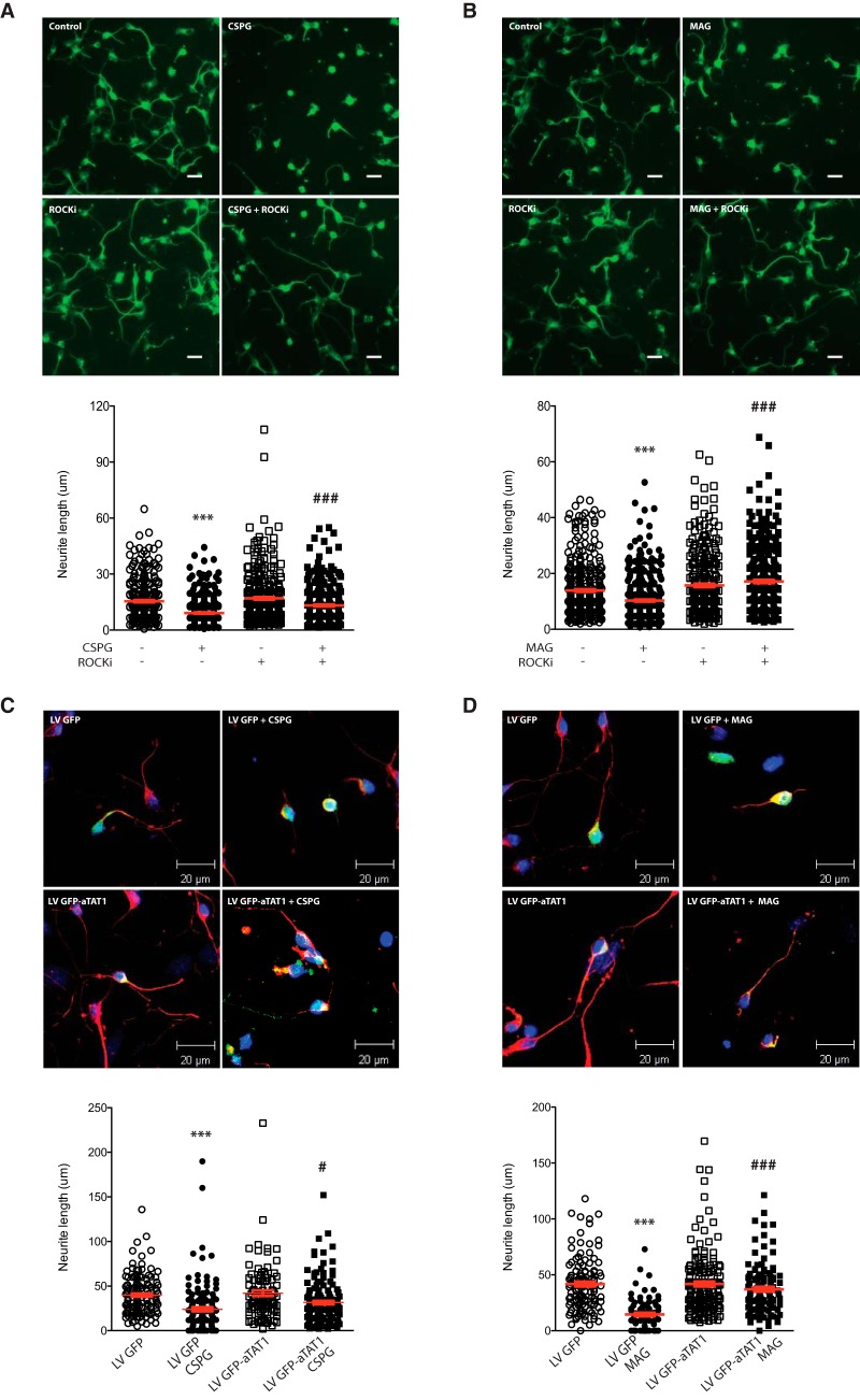
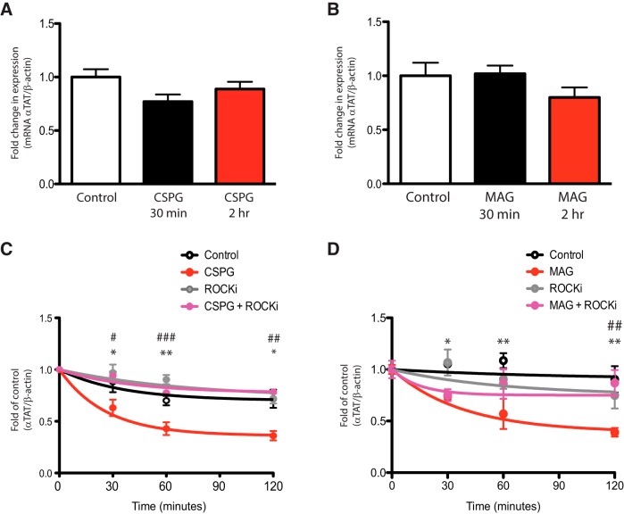
Damage to the CNS results in neuronal and axonal degeneration, and subsequent neurological dysfunction. Endogenous repair in the CNS is impeded by inhibitory chemical and physical barriers, such as chondroitin sulfate proteoglycans (CSPGs) and myelin-associated glycoprotein (MAG), which prevent axon regeneration. Previously, it has been demonstrated that the inhibition of axonal histone deacetylase-6 (HDAC6) can promote microtubule α-tubulin acetylation and restore the growth of CSPGs- and MAG-inhibited axons. Since the acetylation of α-tubulin is regulated by two opposing enzymes, HDAC6 (deacetylation) and α-tubulin acetyltransferase-1 (αTAT1; acetylation), we have investigated the regulation of these enzymes downstream of a growth inhibitory signal. Our findings show that exposure of primary mouse cortical neurons to soluble CSPGs and MAG substrates cause an acute and RhoA-kinase-dependent reduction in α-tubulin acetylation and αTAT1 protein levels, without changes to either HDAC6 levels or HDAC6 activity. The CSPGs- and MAG-induced reduction in αTAT1 occurs primarily in the distal and middle regions of neurites and reconstitution of αTAT1, either by Rho-associated kinase (ROCK) inhibition or lentiviral-mediated αTAT1 overexpression, can restore neurite growth. Lastly, we demonstrate that CSPGs and MAG signaling decreases αTAT1 levels posttranscriptionally via a ROCK-dependent increase in αTAT1 protein turnover. Together, these findings define αTAT1 as a novel potential therapeutic target for ameliorating CNS injury characterized by growth inhibitory substrates that are prohibitive to axonal regeneration.

Keywords: chondroitin sulfate proteoglycan; myelin-associated glycoprotein; α-tubulin acetylation; α-tubulin acetyltransferase.


Figures

None
Graphical abstract
Figure 1.
Figure 1.
Growth inhibitory factors downregulate α-tubulin acetylation and αTAT1 levels. A, B, Immunoblot analysis of primary murine cortical neurons after exposure to soluble CSPGs (2 μg/ml; A) or MAG (30 μg/ml; B) showed a significant decrease in α-tubulin acetylation levels at the indicated times. Acetylated α-tubulin was normalized to total α-tubulin from the same immunoblot. C, D, Immunoblot analysis for HDAC6 after incubation with CSPGs (C) or MAG (D) for 2 h. HDAC6 level was normalized to β-actin from the same immunoblot. E, F, HDAC6 activity assays in primary neurons exposed to CSPGs (E) or MAG (F) after 30 min or 2 h did not change HDAC6 activity. Tubastatin A, a specific HDAC6 inhibitor, was used a positive control. G, H, Immunoblot analysis for αTAT1 after incubation with CSPGs (G) or MAG (H) for 30 min or 2 h showed a signification reduction in αTAT1 protein levels. αTAT1 level was normalized to β-actin from the same immunoblot. *, Significant downregulation compared to the control group p < 0.05; **p < 0.01 (one-way ANOVA followed by Bonferroni’s post hoc test was performed for A, B, E–H. Student’s t test was performed for C, D).
Figure 2.
Figure 2.
Downregulation of αTAT1 and α-tubulin acetylation by CSPGs and MAG is mediated through ROCK-dependent pathway. Primary cortical neurons were treated with either CSPGs (2 μg/ml) or MAG (30 μg/ml) at indicated times, with or without ROCK inhibitor (Y-27632; 10 μM). A, B, Immunoblot analysis for αTAT1 showed that ROCK inhibitor prevented downregulation of αTAT1 after exposure to CSPGs (A) and MAG (B). αTAT1 level was normalized to β-actin from the same immunoblot. C, D, Immunoblot analysis for acetylated α-tubulin showed that ROCK inhibitor also prevented CSPGs- and MAG-induced (C, D, respectively) reduction of α-tubulin acetylation. Acetylated α-tubulin was normalized to total α-tubulin from the same immunoblot. *, Significant downregulation compared to the control group at their respective times, p < 0.05; **p < 0.01 (two-way ANOVA followed by Bonferroni’s post hoc test was performed).
Figure 3.
Figure 3.
CSPGs and MAG change neurite αTAT1 expression. A, D, Confocal immunofluorescent micrographs showing the distribution of αTAT1 in cortical neurons after exposure to growth inhibitory factors CSPGs (2 μg/ml; A) or MAG (30 μg/ml; D) with or without ROCK inhibitor (Y-27632; 10 μM) after 30 min and 2 h. Immunolabeling was performed using antibodies against αTAT1 (1:200; red) and Tuj1 (1:5000; green). Nuclei of neurons were labeled with DAPI (blue). Immunofluorescence intensity at different regions of the axon as indicated by white dashed line (i.e., distal, middle, and NIS) was quantified in B, C and E, F. *, Treatment versus control p < 0.05; **, treatment versus control p < 0.01; ***, treatment versus control p < 0.001; ##, cotreatment with MAG and ROCKi versus MAG alone p < 0.01; ###, cotreatment with MAG and ROCKi versus MAG alone p < 0.001 (two-way ANOVA followed by Bonferroni’s post hoc test was performed). Scale bar, 20 μm.
Figure 4.
Figure 4.
CSPGs and MAG change neurite α-tubulin acetylation. A, D, Confocal immunofluorescent micrographs showing the distribution of αTAT1 in cortical neurons after exposure to growth inhibitory factors CSPGs (2 μg/ml; A) or MAG (30 μg/ml; D) with or without ROCK inhibitor (Y-27632; 10 μM) after 30 min and 2 h. Immunolabeling was performed using antibodies against acetylated α-tubulin (1:1000; red) and α-tubulin (1:5000; green). Nuclei of neurons were labeled with DAPI (blue). Immunofluorescence intensity at different regions of the axon as indicated by white dashed line (i.e., distal, middle, and NIS) was quantified in B, C and E, F. *, Treatment versus control p < 0.05; ***, treatment versus control p < 0.001; #, cotreatment with ROCKi versus treatment alone p < 0.05; ##, cotreatment with ROCKi versus treatment alone p < 0.01 (two-way ANOVA followed by Bonferroni’s post hoc test was performed). Scale bar, 20 μm.
Figure 5.
Figure 5.
ROCK inhibition and overexpression of αTAT1 reverse CSPGs- and MAG-induced inhibition of neurite outgrowth. A, B, Fluorescent microscopy of primary cortical neurons incubated with CSPGs (2 μg/ml; A) or MAG (30 μg/ml; B), with or without ROCK inhibitor (Y-27632; 10 μM) for 24 h. Neurite lengths and mean neurite length for each condition are shown in column scatter plots below micrographs. ***, CSPGs or MAG treatment versus untreated control p < 0.001; ###, cotreatment with ROCKi versus treatment alone p < 0.001 (one-way ANOVA followed by Bonferroni’s post hoc test was performed). Scale bar, 10 μm (A, B). C, D, Confocal immunofluorescent microscopy of primary cortical neurons following infection with lentiviral GFP (LV GFP; control) or lentiviral GFP-αTAT1 (LV GFP-αTAT1) with or without CSPGs (2 μg/ml; C) or MAG (30 μg/ml; D). Transduced neurites were identified by immunolabeling with antibodies for neuron-specific Tuj1 (1:5000; red) and GFP (1:500; green) and quantified with ImageJ software. Neurite lengths and mean neurite length for each condition are shown in column scatter plots below micrographs. ***, CSPGs or MAG treatment versus untreated control p < 0.001; # and ###, LV GFP-αTAT1 with CSPGs or MAG versus LV GFP with CSPGs or MAG, p < 0.05 and p < 0.001, respectively (two-way ANOVA followed by Bonferroni’s post hoc test was performed). Scale bar, 20 μm (C, D).
Figure 6.
Figure 6.
αTAT1 protein stability is reduced in cortical neurons treated with CSPGs or MAG. A, B, Bar graphs showing real-time quantitative RT-PCR results from primary cortical neurons incubated with CSPGs (2 μg/ml; A) or MAG (30 μg/ml; B) for 30 min or 2 h reveal no change in αTAT1 mRNA. C, D, Cycloheximide chase assay graphs showing αTAT1 protein degradation in primary cortical neurons over time after protein translation inhibition with cycloheximide (10 μg/ml). Neurons were treated with or without CSPGs (2 μg/ml; C) or MAG (30 μg/ml; D) and cotreated with or without ROCK inhibitor (Y-27632; 10 μM). *, Treatment with growth inhibitory substrate versus 0 min p < 0.05; **, treatment with growth inhibitory substrate versus 0 min, p < 0.01; #, CSPGs or MAG cotreatment with ROCKi versus CSPGs or MAG treatment alone at 30 min, p < 0.05; ###, CSPGs or MAG cotreatment with ROCKi versus CSPGs or MAG treatment alone at 60 min, p < 0.001; ##, CSPGs or MAG treatment with ROCKi versus CSPGs or MAG treatment alone at 120 min, p < 0.01 (two-way ANOVA followed by Bonferroni’s post hoc test was performed).

References

    1. Akella JS, Wloga D, Kim J, Starostina NG, Lyons-Abbott S, Morrissette NS, Dougan ST, Kipreos ET, Gaertig J (2010) MEC-17 is an alpha-tubulin acetyltransferase. Nature 467:218–222. 10.1038/nature09324 - DOI - PMC - PubMed
    1. Alper JD, Decker F, Agana B, Howard J (2014) The motility of axonemal dynein is regulated by the tubulin code. Biophys J 107:2872–2880. - PMC - PubMed
    1. Baas PW, Ahmad FJ, Pienkowski TP, Brown A, Black MM (1993) Sites of microtubule stabilization for the axon. J Neurosci 13:2177–2185. - PMC - PubMed
    1. Black MM, Baas PW, Humphries S (1989) Dynamics of alpha-tubulin deacetylation in intact neurons. J Neurosci 9:358–368. - PMC - PubMed
    1. Borisoff JF, Chan CC, Hiebert GW, Oschipok L, Robertson GS, Zamboni R, Steeves JD, Tetzlaff W (2003) Suppression of Rho-kinase activity promotes axonal growth on inhibitory CNS substrates. Mol Cell Neurosci 22:405–416. - PubMed

Publication types

LinkOut - more resources