. 2018 Jul 1;315(1):H18-H32.
doi: 10.1152/ajpheart.00683.2017. Epub 2018 Mar 2.

Minoxidil improves vascular compliance, restores cerebral blood flow, and alters extracellular matrix gene expression in a model of chronic vascular stiffness

Affiliations

Minoxidil improves vascular compliance, restores cerebral blood flow, and alters extracellular matrix gene expression in a model of chronic vascular stiffness

Russell H Knutsen et al. Am J Physiol Heart Circ Physiol. .

Abstract

Increased vascular stiffness correlates with a higher risk of cardiovascular complications in aging adults. Elastin (ELN) insufficiency, as observed in patients with Williams-Beuren syndrome or with familial supravalvular aortic stenosis, also increases vascular stiffness and leads to arterial narrowing. We used Eln+/- mice to test the hypothesis that pathologically increased vascular stiffness with concomitant arterial narrowing leads to decreased blood flow to end organs such as the brain. We also hypothesized that drugs that remodel arteries and increase lumen diameter would improve flow. To test these hypotheses, we compared carotid blood flow using ultrasound and cerebral blood flow using MRI-based arterial spin labeling in wild-type (WT) and Eln+/- mice. We then studied how minoxidil, an ATP-sensitive K+ channel opener and vasodilator, affects vessel mechanics, blood flow, and gene expression. Both carotid and cerebral blood flows were lower in Eln+/- mice than in WT mice. Treatment of Eln+/- mice with minoxidil lowered blood pressure and reduced functional arterial stiffness to WT levels. Minoxidil also improved arterial diameter and restored carotid and cerebral blood flows in Eln+/- mice. The beneficial effects persisted for weeks after drug removal. RNA-Seq analysis revealed differential expression of 127 extracellular matrix-related genes among the treatment groups. These results indicate that ELN insufficiency impairs end-organ perfusion, which may contribute to the increased cardiovascular risk. Minoxidil, despite lowering blood pressure, improves end-organ perfusion. Changes in matrix gene expression and persistence of treatment effects after drug withdrawal suggest arterial remodeling. Such remodeling may benefit patients with genetic or age-dependent ELN insufficiency. NEW & NOTEWORTHY Our work with a model of chronic vascular stiffness, the elastin ( Eln)+/- mouse, shows reduced brain perfusion as measured by carotid ultrasound and MRI arterial spin labeling. Vessel caliber, functional stiffness, and blood flow improved with minoxidil. The ATP-sensitive K+ channel opener increased Eln gene expression and altered 126 other matrix-associated genes.

Keywords: ATP-sensitive K+ channel; arterial stiffness; cerebral blood flow; elastin; extracellular matrix; vascular remodeling.


Figures
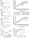
Fig. 1.
Fig. 1.
Minoxidil decreases blood pressure (BP) and increases vessel diameter in elastin (Eln)+/− mice. Mice received minoxidil in drinking water from weaning to 3 mo of age. A–C: systolic BP (SBP; A), diastolic BP (DBP; B), and pulse pressure (C; by one-way ANOVA). D and E: pressure-diameter curves in the aorta (D) and carotid (E) arteries (two-way repeated-measures ANOVA). F and G: aortic segmental distensibility (SD25; F) and functional distensibility (FD; G) at average BP (by two-way repeated-measures ANOVA). In A−G, wild-type (WT) mice are shown as black circles and Eln+/− mice are shown as gray squares. Minoxidil-treated Eln+/− mice are shown as open squares. Means and standard deviations are shown as whiskers underlying the data points. On the pressure-diameter curves, untreated animals [WT (black) and Eln+/− (gray)] are shown as solid lines and minoxidil-treated Eln+/− animals are shown as dashed lines. Comparisons (Tukey’s) among individual groups are shown. ^^Minoxidil-treated Eln+/− vs. untreated WT comparisons; **minoxidil-treated Eln+/− vs. untreated Eln+/− comparisons; ##untreated WT vs. untreated Eln+/− tests; *,^P < 0.05, **,^^,##P < 0.01, ***,^^^,###P < 0.001, and ****,^^^^,####P < 0.0001. Treatment with minoxidil reduced SBP and pulse pressure and increased aortic and carotid diameters at multiple pressures. SD25 in the aorta of minoxidil-treated Eln+/− mice was similar to untreated Eln+/− mice, but FD was intermediate. The remaining comparisons were not significant. The gray box represents the average aortic BP range of minoxidil-treated Eln+/− mice (112/74 mmHg). OD, outer diameter.
Fig. 2.
Fig. 2.
Minoxidil increases elastin (ELN) but not collagen in Eln+/− aortas. Mice received minoxidil in drinking water from weaning to 3 mo of age. Desmosine (ELN; A and B) and hydroxyproline (collagen; C and D) content are shown relative to the ascending aortic segment (A and C) and total protein (B and D). Genotype and treatment status are shown below each graph (one-way ANOVA). Multiple comparisons (Tukey’s) are shown. *P < 0.05; **P < 0.01; ***P < 0.001; ****P < 0.0001. Desmosine was higher in wild-type (WT) untreated and minoxidil-treated Eln+/− mice than in untreated Eln+/− mice regardless of normalization, although the differences were reduced when normalized to total protein. Collagen content, as assessed by hydroxyproline level, showed no differences among the groups tested. Means and standard deviations are shown by error bars.
Fig. 3.
Fig. 3.
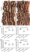
Elastin (ELN) changes in minoxidil-treated vessels. A−D: histological sections of unloaded descending aortas from untreated wild-type (WT; A), untreated Eln+/− (B), minoxidil-treated WT (C), and minoxidil-treated Eln+/−(D) mice are shown stained with elastin van Gieson stain. ELN is dark. E: lamellar number for the descending aorta. F: total area that stained with the ELN stain. G: ELN-stained area as a fraction of the total wall area. H: per lamella ELN area. Genotype and treatment status are shown below each graph (one-way ANOVA). Multiple comparisons (Tukey’s) are shown. *P < 0.05; **P < 0.01; ***P < 0.001; ****P < 0.0001. All other comparisons were not significant. Means and standard deviations are shown by error bars.
Fig. 4.
Fig. 4.
Minoxidil induces vessel remodeling and growth. A: lumen area from wild-type (WT), untreated elastin (Eln)+/−, and minoxidil-treated Eln+/− mice. B: wall thickness. C: wall cross-sectional area. D: wall cross-sectional-to-lumen area ratio. Genotype and treatment status are shown below each graph (one-way ANOVA). Multiple comparisons (Tukey’s) are shown. *P < 0.05; **P < 0.01; ***P < 0.001; ****P < 0.0001. All other comparisons were not significant. Means and standard deviations are shown by error bars.
Fig. 5.
Fig. 5.
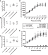
Vessels from elastin (Eln)+/− mice removed from minoxidil remain larger than those from untreated animals. Eln+/− mice received minoxidil in drinking water from weaning to 3 mo, as shown in Fig. 1. Additional mice received the drug until 3 mo and water only (no drug) from ages of 3 to 4 mo (Eln+/− minoxidil on/off). Mice were euthanized at 4 mo along with untreated 4-mo control mice. A−C: systolic blood pressure (BP; A), diastolic BP (B), and pulse pressure (C; by two-way ANOVA). D and E: pressure-diameter curves in the aorta (D) and carotid (E) arteries (by two-way repeated-measures ANOVA). 3-mo-old Eln+/−mice are shown by gray symbols, 4-mo-old Eln+/− mice are shown as black symbols; untreated animals are shown as solid lines, and minoxidil-treated animals are shown by dashed lines and open symbols. Multiple testing results (Dunnett’s) comparing the 4-mo Eln+/− minoxidil on/off group to the remaining three groups are shown. D and E: P values comparing 4-mo Eln+/− minoxidil on/off mice with 3-mo minoxidil-treated mice are denoted as ^^; P values with 3-mo untreated mice are denoted as ##. P values comparing 4-mo minoxidil on/off Eln+/− and 4-mo untreated Eln+/− mice are denoted as **. * (or ^ or #) P < 0.05, ** (or ^^ or ##) P < 0.01, and *** (or ^^^ or ###) P < 0.001. The remaining comparisons were not significant. While BP reverted to untreated levels off drug, 4-mo-old treated then off Eln+/− mice trended between treated 3-mo-old Eln+/− and untreated mice, with varying degrees of significance. Means and standard deviations are shown by error bars.
Fig. 6.
Fig. 6.
Minoxidil normalizes in vivo diameter, pulse wave velocity, and carotid blood flow in chronically treated elastin (Eln)+/− mice. Mice received minoxidil in drinking water from weaning to 3 mo of age. A−C: in vivo lumen diameter (systolic and diastolic; A), pulse wave velocity (B), and carotid artery Doppler flow (C) for wild-type (WT) mice (black circles) and Eln+/− mice (gray squares) treated with water (closed symbols) or minoxidil (open symbols). One-way ANOVA and Tukey’s multiple comparisons are shown. *P < 0.05, **P < 0.01, ***P < 0.001, and ****P < 0.0001. In each case, minoxidil treatment returned abnormal Eln+/− values to near WT levels. D: carotid flow for all vessels tested as a function of carotid radius to the fourth power (R4) at 100 mmHg. Means and standard deviations are shown by error bars in AC.
Fig. 7.
Fig. 7.
Elastin (ELN) insufficiency decreases brain perfusion in mice. A and B: representative perfusion maps for wild-type (WT) and Eln+/− cohorts (A) along with plots of arterial spin-labeling MRI-measured perfusion in the whole brain, cortex, hippocampus, and thalamus regions of interest (ROIs; B). Perfusion maps suggested reduced global perfusion in the Eln+/− cohort (light gray) compared with the WT cohort (black), particularly in the cortex, where perfusion was reduced from 251 ml·100 g−1·min−1 in WT mice to 209 ml·100 g−1·min−1 in Eln+/− mice (P < 0.05 by t-test). No group differences were seen in the whole brain, hippocampal, and thalamic ROI analyses, although the perfusion in the Eln+/− cohort trended lower in these regions compared with the WT cohort. For each t-test, *P < 0.05. Means and standard deviations are also shown.
Fig. 8.
Fig. 8.
Minoxidil increases cortical perfusion in elastin (Eln)+/− mice. Mice were treated from weaning to 3 mo of age with minoxidil in drinking water. Minoxidil treatment increased cortical perfusion in Eln+/− mice. Cortical perfusion increased from 209 ml·100 g−1·min−1 in untreated Eln+/− mice (gray filled squaress) to 250 ml·100 g−1·min−1 in minoxidil-treated Eln+/− mice (gray open squares). *P < 0.05 by t-test. Means and standard deviations are also shown.
Fig. 9.
Fig. 9.
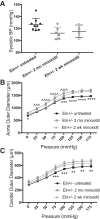
Minoxidil treatment of elastin (Eln)+/− mice for 2 wk shows vessel size intermediate between untreated and 2-mo-treated mice. Eln+/− mice received minoxidil in drinking water for 2 wk or 2 mo. Untreated mice are also shown. A: systolic blood pressure (BP). B and C: pressure-diameter curves in the aorta (B) and carotid (C). Untreated Eln+/− mice and Eln+/− mice treated with minoxidil for 2 mo are shown using black or gray solid lines, respectively, and filled boxes. These are the same data used in Fig. 1. Four mice treated with minoxidil for 2 wk are shown using gray dashed lines and open gray boxes. Means and standard deviations are shown as whiskers underlying the data points. For A–C, one-way ANOVA demonstrated P < 0.05 or better. Dunnett’s multiple comparisons were performed comparing the 2-wk treated Eln+/− group with the other treatment groups. P values comparing 2-wk Eln+/− minoxidil-treated with 2-mo-minoxidil-treated mice are denoted with ^, and P values with untreated mice are denoted with *. *P < 0.05, **P < 0.01, ***,^^^P < 0.001, and ****,^^^^P < 0.0001. The remaining comparisons were not significant.
Fig. 10.
Fig. 10.
Differentially expressed genes with minoxidil treatment and discontinuation. A: Venn diagram with the numbers of differentially expressed genes for each comparison. All differentially expressed genes with expression differences of at least 1.5-fold with a false discovery rate of 0.1 between any two of the treatment groups were plotted by heat map. The four main subgroups [clusters 1−4 (C1–4)] are shown in B. C: log2 mean-centered expression of genes within a given cluster in untreated elastin (Eln)+/− mice, mice treated with minoxidil for 2 wk, and mice treated for 2 wk and then removed from the drug for 1 wk. The expression profiles for Eln and lysyl oxidase (Lox) are shown superimposed on the cluster data (dashed lines).

Similar articles

Cited by

References

    1. Carta L, Wagenseil JE, Knutsen RH, Mariko B, Faury G, Davis EC, Starcher B, Mecham RP, Ramirez F. Discrete contributions of elastic fiber components to arterial development and mechanical compliance. Arterioscler Thromb Vasc Biol 29: 2083–2089, 2009. doi:10.1161/ATVBAHA.109.193227. - DOI - PMC - PubMed
    1. Cherniske EM, Carpenter TO, Klaiman C, Young E, Bregman J, Insogna K, Schultz RT, Pober BR. Multisystem study of 20 older adults with Williams syndrome. Am J Med Genet A 131: 255–264, 2004. doi:10.1002/ajmg.a.30400. - DOI - PubMed
    1. Coquand-Gandit M, Jacob MP, Fhayli W, Romero B, Georgieva M, Bouillot S, Estève E, Andrieu JP, Brasseur S, Bouyon S, Garcia-Honduvilla N, Huber P, Buján J, Atanasova M, Faury G. Chronic treatment with minoxidil induces elastic fiber neosynthesis and functional improvement in the aorta of aged mice. Rejuvenation Res 20: 218–230, 2017. doi:10.1089/rej.2016.1874. - DOI - PubMed
    1. Davies M, Udwin O, Howlin P. Adults with Williams syndrome. Preliminary study of social, emotional and behavioural difficulties. Br J Psychiatry 172: 273–276, 1998. doi:10.1192/bjp.172.3.273. - DOI - PubMed
    1. Dobin A, Davis CA, Schlesinger F, Drenkow J, Zaleski C, Jha S, Batut P, Chaisson M, Gingeras TR. STAR: ultrafast universal RNA-seq aligner. Bioinformatics 29: 15–21, 2013. doi:10.1093/bioinformatics/bts635. - DOI - PMC - PubMed

Publication types

MeSH terms