3D printing and intraoperative neuronavigation tailoring for skull base reconstruction after extended endoscopic endonasal surgery: proof of concept
- PMID: 29498576
- PMCID: PMC6119650
- DOI: 10.3171/2017.9.JNS171253
3D printing and intraoperative neuronavigation tailoring for skull base reconstruction after extended endoscopic endonasal surgery: proof of concept
Abstract
Objective: Endoscopic endonasal approaches are increasingly performed for the surgical treatment of multiple skull base pathologies. Preventing postoperative CSF leaks remains a major challenge, particularly in extended approaches. In this study, the authors assessed the potential use of modern multimaterial 3D printing and neuronavigation to help model these extended defects and develop specifically tailored prostheses for reconstructive purposes.
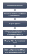
Methods: Extended endoscopic endonasal skull base approaches were performed on 3 human cadaveric heads. Pre-Preprocedure and intraprocedure CT scans were completed and were used to segment and design extended and tailored skull base models. Multimaterial models with different core/edge interfaces were 3D printed for implantation trials. A novel application of the intraoperative landmark acquisition method was used to transfer the navigation, helping to tailor the extended models.
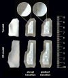
Results: Prostheses were created based on preoperative and intraoperative CT scans. The navigation transfer offered sufficiently accurate data to tailor the preprinted extended skull base defect prostheses. Successful implantation of the skull base prostheses was achieved in all specimens. The progressive flexibility gradient of the models’ edges offered the best compromise for easy intranasal maneuverability, anchoring, and structural stability. Prostheses printed based on intraprocedure CT scans were accurate in shape but slightly undersized.
Conclusions: Preoperative 3D printing of patient-specific skull base models is achievable for extended endoscopic endonasal surgery. The careful spatial modeling and the use of a flexibility gradient in the design helped achieve the most stable reconstruction. Neuronavigation can help tailor preprinted prostheses.
Keywords: 3D; endoscopic; extended endonasal surgery; neuronavigation; printing; prostheses and implants; reconstructive surgical procedures; skull base reconstruction; surgical technique.
Conflict of interest statement
The authors report no conflict of interest concerning the materials or methods used in this study or the findings specified in this paper.
Figures


References
-
- Ahmed OH, Marcus S, Tauber JR, Wang B, Fang Y, Lebowitz RA. Efficacy of perioperative lumbar drainage following endonasal endoscopic cerebrospinal fluid leak repair: a meta-analysis. Otolaryngol Head Neck Surg. 2017;156:52–60. - PubMed
-
- Alvarez Berastegui GR, Raza SM, Anand VK, Schwartz TH. Endonasal endoscopic transsphenoidal chiasmapexy using a clival cranial base cranioplasty for visual loss from massive empty sella following macroprolactinoma treatment with bromocriptine: case report. J Neurosurg. 2016;124:1025–1031. - PubMed
-
- Bandyopadhyay A, Bose S, Das S. 3D printing of biomaterials. MRS Bull. 2015;40:108–115.
-
- Banu MA, Kim JH, Shin BJ, Woodworth GF, Anand VK, Schwartz TH. Low-dose intrathecal fluorescein and etiology-based graft choice in endoscopic endonasal closure of CSF leaks. Clin Neurol Neurosurg. 2014;116:28–34. - PubMed
-
- Bartlett NW, Tolley MT, Overvelde JT, Weaver JC, Mosadegh B, Bertoldi K, et al. A 3D-printed, functionally graded soft robot powered by combustion. Science. 2015;349:161–165. - PubMed
Publication types
MeSH terms
Grants and funding
LinkOut - more resources
Full Text Sources
Other Literature Sources
Medical
Molecular Biology Databases

