. 2018 Mar 2;23(3):564.
doi: 10.3390/molecules23030564.

Synergistic Effects of Salvianolic Acid B and Puerarin on Cerebral Ischemia Reperfusion Injury

Affiliations

Synergistic Effects of Salvianolic Acid B and Puerarin on Cerebral Ischemia Reperfusion Injury

Chengli Ling et al. Molecules. .

Abstract

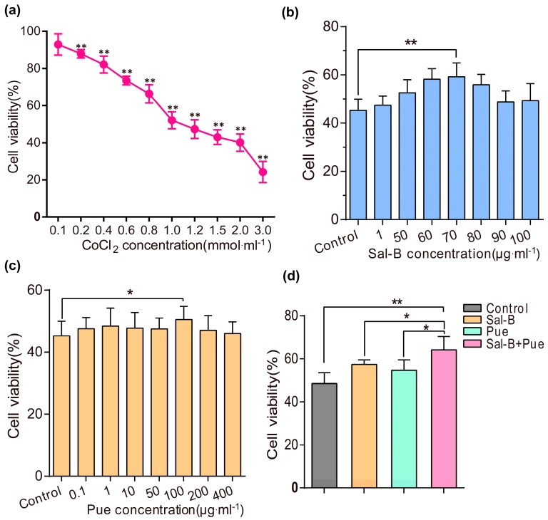
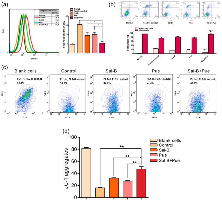
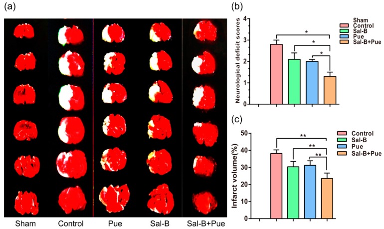
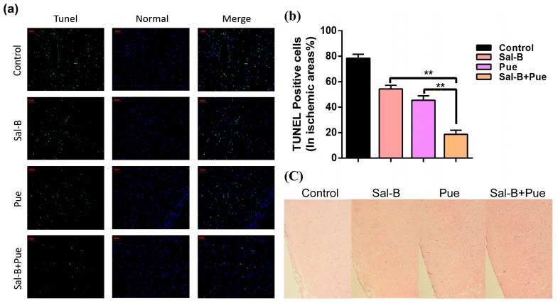
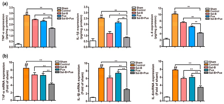
Ischemic stroke (IS) is characterized by the sudden loss of blood circulation to an area of the brain, resulting in a corresponding loss of neurologic function. It has been a worldwide critical disease threatening to the health and life of human beings. Despite significant progresses achieved, effective treatment still remains a formidable challenge due to the complexity of the disease. Salvianolic acid B (Sal-B) and Puerarin (Pue) are two active neuroprotectants isolated from traditional Chinese herbs, Salvia miltiorrhiza and Kudzu root respectively, which have been used for the prevention and treatment of IS for thousands of years in China. The activities of two compounds against cerebral ischemia reperfusion injury have been confirmed via various pathways. However, the therapeutic efficacy of any of the two components is still unsatisfied. In the present study, the effect of the combination of Sal-B and Pue on IS was evaluated and validated in vitro and in vivo. The ratio of two compounds was firstly optimized based on the results of CoCl₂ damaged PC12 cells model. The co-administration exhibited significantly protective effect in CoCl₂ induced PC12 cells injury model by reducing ROS, inhibiting apoptosis and improving mitochondrial membrane potential in vitro. Moreover, Sal-B + Pue significantly relieved neurological deficit scores and infarct area than Sal-B or Pue alone in vivo. The results indicated that neuroprotection mechanism of Sal-B + Pue was related to TLR4/MyD88 and SIRT1 activation signaling pathway to achieve synergistic effect, due to the inhibition of NF-κB transcriptional activity and expression of pro-inflammatory cytokine (TNF-α, IL-1β, IL-6). In conclusion, the combination of Sal-B and Pue exerted much stronger neuroprotective effect than Sal-B or Pue alone, which provides a potential new drug and has great significance for the treatment of IS.

Keywords: Salvianolic acid B; combination; inflammation; ischemia stroke; puerarin; synergistic effect.


Conflict of interest statement

The authors declare no conflict of interest.

Figures

Figure 1
Figure 1
The chemical structures of Sal-B and Pue.
Figure 2
Figure 2
Protective effects of Sal-B, Pue and Sal-B + Pue in CoCl2 injured PC12 cells. (a) The viability of PC12 cells induced with different concentration (0.1, 0.2, 0.4, 0.6, 0.8, 1, 1.2, 1.5, 2, 3 mmol·L−1) of CoCl2 for 24 h (** p < 0.01 vs. control group); (b) The viability of PC12 cells pretreated with various concentrations of Sal-B (1, 50, 60, 70, 80, 90, 100 μg·mL−1) and (c) Pue (0.1, 1, 10, 50, 100, 200, 400 μg·mL−1) for 1 h before induced by CoCl2 (* p < 0.05; ** p < 0.01 vs. control); (d) The viability of PC12 cells pretreated with Sal-B (70 μg·mL−1) and Pue (100 μg·mL−1) simultaneously for 1 h before induced by CoCl2. Data were expressed as mean ± SD (n = 6), * p < 0.05 vs. Sal-B or Pue; ** p < 0.01 Sal-B + Pue vs. blank control.
Figure 3
Figure 3
Effect of the combination of Sal-B and Pue on (a) ROS level; (b) apoptosis; (c) mitochondrial membrane potential on CoCl2 injured PC12 cells and (d) quantitative statistic result of JC-1 aggregates, representing the fluctuation of MMP. Data were expressed as mean ± SD (n = 3), * ≠Γp < 0.05 Sal-B + Pue vs. positive control, Sal-B or Pue in apoptosis; * p < 0.05; ** p < 0.01 vs. other group.
Figure 4
Figure 4
(a) Effects of Sal-B, Pue, Sal-B + Pue on infarct volume and neurological functional outcome (n = 6). (TTC staining) White area indicates infarcted brain tissue; (b) Neurological deficit score; (c) Percentage of cerebral infarction volumes were calculated as described in “Methods.” The Sal-B + Pue group exhibited obviously less cerebral infraction (white area) compared with other groups. * p < 0.05; ** p < 0.01 vs. other group.
Figure 5
Figure 5
(a) TUNEL and (c) H&E microscopic images of brain sections 24 h after drug treatment. The green indicates the apoptotic cells and the blue represents normal cells in ischemic area (200 μm). The damaged neurons with apoptotic and necrotic changes were labeled (brown) at H&E staining and original magnification was 200×. (b) Quantitative results of apoptosis in ischemia area (n = 3), ** p < 0.01 vs. Sal-B or Pue.
Figure 6
Figure 6
Effect of Sal-B, Pue and Sal-B + Pue treatment on (a) the protein expression and (b) mRNA levels of pro-inflammatory mediators TNF-α, IL-1β, IL-6 in the penumbra of the brain after 1.5 h MCAO and 24 h reperfusion injury (n = 6). The values were β-actin normalized and the relative mRNA levels in the sham-operated group were used as calibrators. Data are expressed as the mean ± SD (## p < 0.01 vs. sham; * p < 0.05 and ** p < 0.01 vs. control, Sal-B or Pue).
Figure 7
Figure 7
(a) Effects of Sal-B, Pue and Sal-B + Pue on the protein levels of TLR4, MyD88, SIRT1 and NF-κB in the brain tissue of penumbra (n = 3) after 1.5 h MCAO and 24 h reperfusion injury and (b) the statistical analysis results. Data are presented as mean ± SD. (## p < 0.01 each group vs. control group, “ns” represents non-significant difference; * p < 0.05; ** p < 0.01 vs. Sal-B or Pue).
Figure 8
Figure 8
Schematic diagram for the neuroprotective effects of Sal-B + Pue in I/R injury. Sal-B + Pue suppressed TLR4/MyD88 signaling and SIRT1 activation to reach a synergistic effect after I/R injury.

References

    1. Zhang Q., Zhao Y.H. Therapeutic Angiogenesis after ischemic stroke: Chinese medicines, Bone marrow stromal cells (BMSCs) and their combinational treatment. Am. J. Chin. Med. 2014;42:61–77. doi: 10.1142/S0192415X14500049. - DOI - PubMed
    1. Sahota P., Savitz S.I. Investigational therapies for ischemic stroke: Neuroprotection and neurorecovery. Neurotherapeutics. 2011;8:434–451. doi: 10.1007/s13311-011-0040-6. - DOI - PMC - PubMed
    1. Mattiasson G., Shamloo M., Gido G., Mathi K., Tomasevic G., Yi S., Warden C.H., Castilho R.F., Melcher T., Gonzalez-Zulueta M., et al. Uncoupling protein-2 prevents neuronal death and diminishes brain dysfunction after stroke and brain trauma. Nat. Med. 2003;9:1062–1068. doi: 10.1038/nm903. - DOI - PubMed
    1. Gong X., Sucher N.J. Stroke therapy in traditional Chinese medicine (TCM): Prospects for drug discovery and development. Phytomedcine. 2002;9:478–484. doi: 10.1078/09447110260571760. - DOI - PubMed
    1. Wang F.R., Yang X.W., Zhang Y., Liu J.-X., Yang X.-B., Liu Y., Shi R.-B. Three new isoflavone glycosides from Tongmai granules. J. Asian Nat. Prod. Res. 2011;13:319–327. doi: 10.1080/10286020.2011.556090. - DOI - PubMed

MeSH terms