Review
. 2018 May 21;47(10):3406-3420.
doi: 10.1039/c7cs00827a.

Minimalistic peptide supramolecular co-assembly: expanding the conformational space for nanotechnology

Affiliations
Review

Minimalistic peptide supramolecular co-assembly: expanding the conformational space for nanotechnology

Pandeeswar Makam et al. Chem Soc Rev. .

Abstract

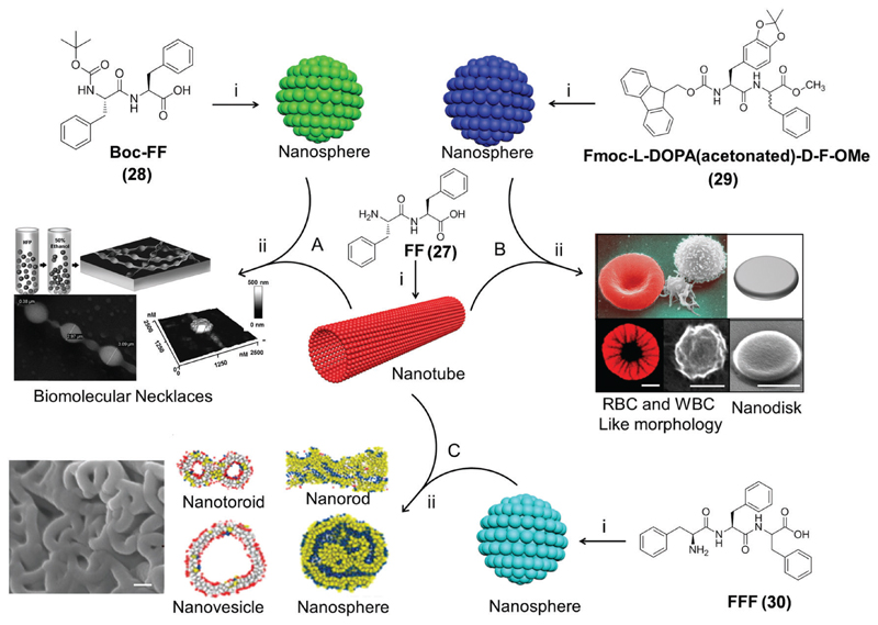
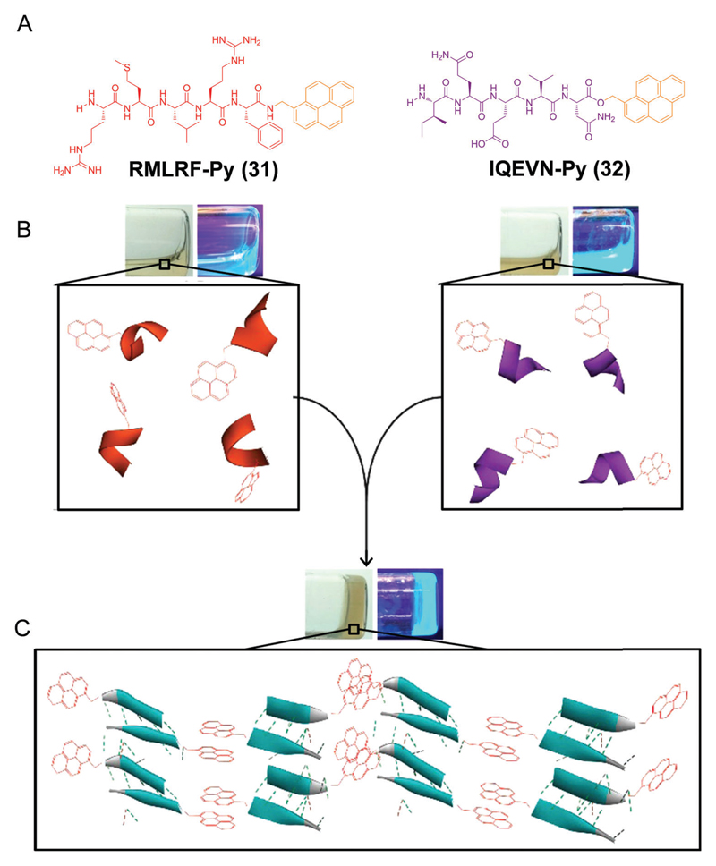
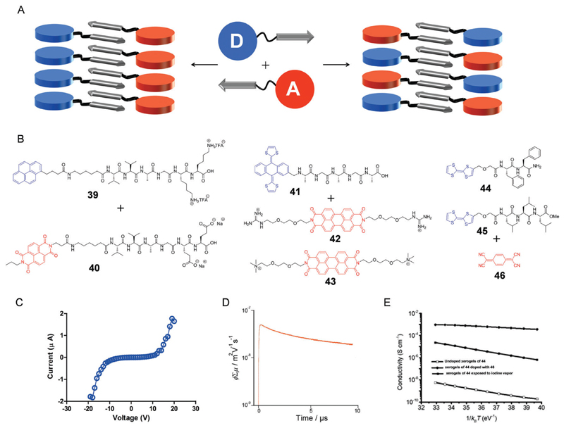
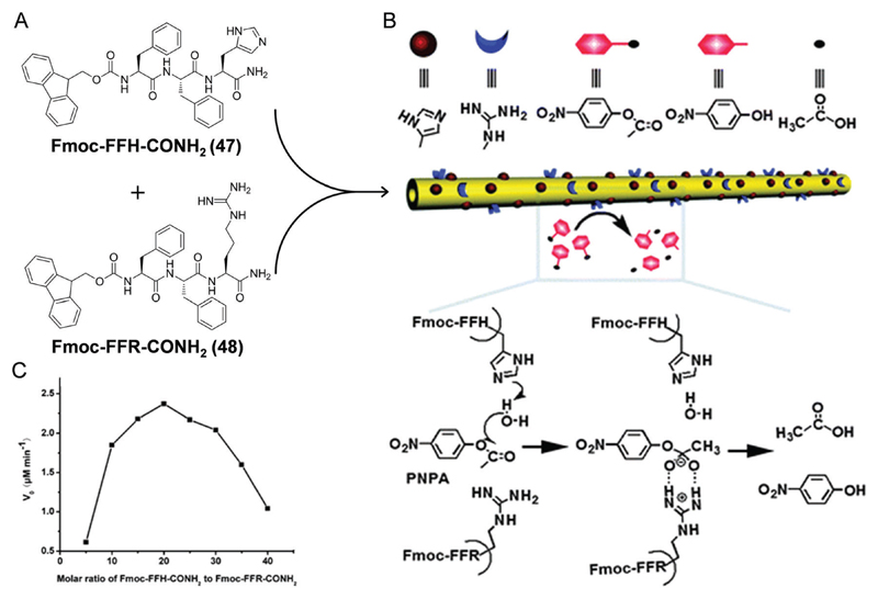
Molecular self-assembly is a ubiquitous process in nature and central to bottom-up nanotechnology. In particular, the organization of peptide building blocks into ordered supramolecular structures has gained much interest due to the unique properties of the products, including biocompatibility, chemical and structural diversity, robustness and ease of large-scale synthesis. In addition, peptides, as short as dipeptides, contain all the molecular information needed to spontaneously form well-ordered structures at both the nano- and the micro-scale. Therefore, peptide supramolecular assembly has been effectively utilized to produce novel materials with tailored properties for various applications in the fields of material science, engineering, medicine, and biology. To further expand the conformational space of peptide assemblies in terms of structural and functional complexity, multicomponent (two or more) peptide supramolecular co-assembly has recently evolved as a promising extended approach, similar to the structural diversity of natural sequence-defined biopolymers (proteins) as well as of synthetic covalent co-polymers. The use of this methodology was recently demonstrated in various applications, such as nanostructure physical dimension control, the creation of non-canonical complex topologies, mechanical strength modulation, the design of light harvesting soft materials, fabrication of electrically conducting devices, induced fluorescence, enzymatic catalysis and tissue engineering. In light of these significant advancements in the field of peptide supramolecular co-assembly in the last few years, in this tutorial review, we provide an updated overview and future prospects of this emerging subject.


Conflict of interest statement

Conflicts of interest

There are no conflicts to declare.

Figures

Fig. 1
Fig. 1
Peptide supramolecular co-polymers. Schematic illustration of how two peptide building blocks (blue and red bricks) can self-assemble into an ordered architecture (blue or red wall); mixing them results in a complex co-assembled architecture (wall comprises both blue and red bricks) via four possible supramolecular co-polymer arrangements such as cooperative, orthogonal (or self-sorting), random and disruptive co-assembly.
Fig. 2
Fig. 2
Peptide supramolecular co-assembly driven dimensional control. (A and B) Chemical structures and schematic illustration of FF nanotube physical dimensional control via co-assembly with Boc-FF at the different molar ratios. (C and D) The corresponding histograms of the nanotube length distributions (μm) (insets: transmission electron microscopy images, scale bar 10 μm) and ToF-SIMS analysis (optical micrograph (left), ToF-SIMS chemical maps (right)) respectively. Reproduced with permission from ref. Copyright 2016, American Chemical Society.
Fig. 3
Fig. 3
Creation of non-canonical complex peptide topologies via supramolecular co-assembly. (A) Co-assembly of FF and Boc-FF into a biomolecular necklace like architecture; scanning electron microscopy (SEM) and 3D atomic force microscopy (AFM) image of co-assembled necklaces (reproduced with permission from ref. Copyright 2012, American Chemical Society). (B) Co-assembly of FF with Fmoc-(l)DOPA(acetonated)-(d)F-OMe into red (RBC) and white (WBC) blood cell-like and disk-like nanostructures, fluorescence (doxorubicin adsorbed) and SEM images, scale bar 1 μm (reproduced with permission from ref. Copyright 2014, Royal Society of Chemistry). (C) Co-assembly of FF with FFF to form diverse molar ratio dependent tunable nanomorphologies including nanotoroids, nanovesicles, nanorods, and nanospheres; SEM image of a nanotoroid, scale bar 100 nm (reproduced with permission from ref. Copyright 2016, American Chemical Society). (i) Self-assembly; (ii) co-assembly process.
Fig. 4
Fig. 4
Co-assembly driven peptide secondary structural transition. (A) Chemical structures of pyrene conjugated peptides. (B) Solutions (under visible and UV light) of individual peptide conjugates comprising an α-helix like conformation. (C) Supramolecular co-assembly driven hydrogels comprising a β-sheet-like conformation. Reproduced with permission from ref. Copyright 2017, American Chemical Society.
Fig. 5
Fig. 5
Light harvesting peptide supramolecular co-assemblies. (A) Schematic illustration of the organic fluorophore (donors (blue disk) and acceptors (red disks)) conjugated peptide component (arrow). Supramolecular co-assembly driven donor–acceptor light harvesting system via (a) absorption of a photon by the donor, (b) nonradiative transfer to an acceptor via resonance energy transfer (RET) and (c) the release of energy from the acceptor (reproduced with permission from ref. Copyright 2009, American Chemical Society). (B) Chemical structures of donor and acceptor peptide conjugate pairs (33/34; 35/36 and 37/38).
Fig. 6
Fig. 6
Electrically conducting peptide supramolecular co-assemblies. (A) Schematic illustration of the organic aromatic chromophore (donors (blue disk) and acceptors (red disks)) conjugated peptide component (arrow) and its possible co-assembly organization. (B) Chemical structures of donor and acceptor peptide conjugate pairs (39/40; 41/42, 41/43, 44/46 and 45/46). (C) Current–voltage (IV) characteristics of the 39/40 co-assembled film (reproduced with permission from ref. Copyright 2017, American Chemical Society). (D) Conductivity transients observed for 41/42 upon exposure at 355 nm, 9.1 x 10¹⁵ photons per cm2 (reproduced with permission from ref. Copyright 2015, American Chemical Society). (E) The temperature-dependent conductivity measurements of the 44 xerogels before and after co-assembly or doping (reproduced with permission from ref. Copyright 2014, American Chemical Society).
Fig. 7
Fig. 7
Biocatalytic supramolecular peptide co-assembly. (A) Molecular structures of peptide amphiphiles. (B) Schematic representation of the coassembly of 47 and 48 into nanotubes with catalytically active functionalities (imidazolyl and guanidyl) and on the surface and possible mechanism for the p-NPA hydrolysis. (C) The plot of catalytic reaction rate vs. molar ratio of Fmoc-FFH-CONH2 to Fmoc-FFR-CONH2. Reproduced with permission from ref. Copyright 2013, Royal Society of Chemistry.
Fig. 8
Fig. 8
Peptide supramolecular co-assemblies as scaffolds for 3D cell culture. (A) Chemical structures of peptide amphiphiles. (B) TEM images of Fmoc-FF/RGD co-assembled nanofibers and the proposed supramolecular co-assembly model. (C) Human adult dermal fibroblast (HDFa) adhesion and morphology in the Fmoc-FF/RGD hydrogels 48 h post culture. (D) Influence of Fmoc-RGD/FF concentration ratio on cell spreading. (E) Cell proliferation in the Fmoc-FF/RGD and Fmoc-FF/RGE hydrogels. Reproduced with permission from ref. Copyright 2009, Elsevier.
Fig. 9
Fig. 9
Peptide supramolecular co-assembly driven fluorescence modulation. (A) Chemical structures of the β-sheet fibrillizing fusion domain and β-sheet fibrillizing peptide. (B) Schematic representation of co-assembly between fusion fluorescent proteins having a β-sheet fibrillizing domain and β-sheet fibrillizing peptides to form nanofibres with a precise combination of protein ligands. (C) Different fluorescent β-Tail proteins co-assembled into microgels at a precisely tunable dose, demonstrating close correlation between the actual gel color and the predicted color, scale bar 40 μm. Reproduced with permission from ref. Copyright 2014, Nature Publishing Group.

References

    1. Lehn J-M. Science. 1993;260:1762–1763. - PubMed
    1. Whitesides GM. Science. 2002;295:2418–2421. - PubMed
    1. Aida T, Meijer EW, Stupp SI. Science. 2012;335:813–817. - PMC - PubMed
    1. Philp D, Stoddart JF. Angew Chem, Int Ed Engl. 1996;35:1154–1196.
    1. Tao K, Makam P, Aizen R, Gazit E. Science. 2017;358:eaam9756. - PMC - PubMed

LinkOut - more resources