Review
. 2018 May 20:47:201-222.
doi: 10.1146/annurev-biophys-070816-033712. Epub 2018 Mar 2.

Structure and Dynamics of Membrane Proteins from Solid-State NMR

Affiliations
Review

Structure and Dynamics of Membrane Proteins from Solid-State NMR

Venkata S Mandala et al. Annu Rev Biophys. .

Abstract

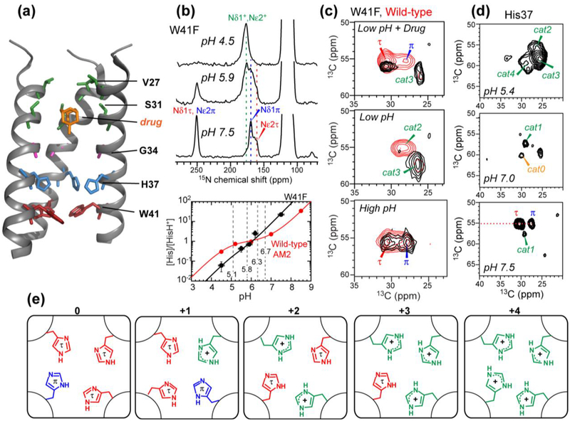
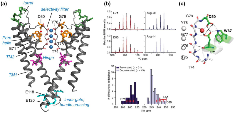
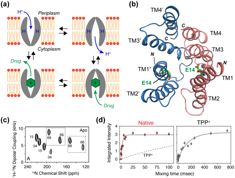
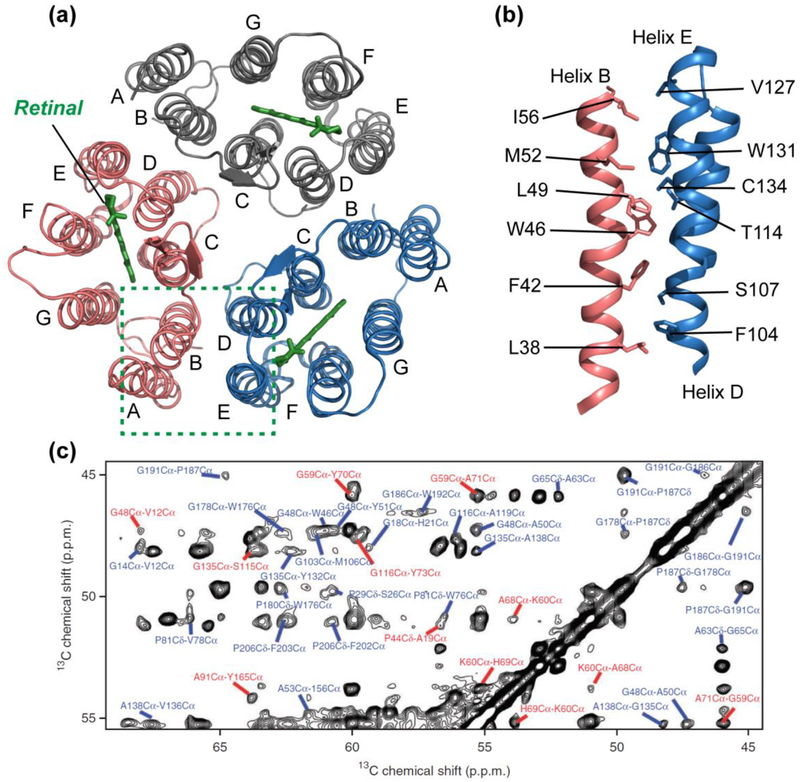
Solid-state nuclear magnetic resonance (SSNMR) spectroscopy elucidates membrane protein structures and dynamics in atomic detail to yield mechanistic insights. By interrogating membrane proteins in phospholipid bilayers that closely resemble biological membranes, SSNMR spectroscopists have revealed ion conduction mechanisms, substrate transport dynamics, and oligomeric interfaces of seven-transmembrane helix proteins. Research has also identified conformational plasticity underlying virus-cell membrane fusions by complex protein machineries, and β-sheet folding and assembly by amyloidogenic proteins bound to lipid membranes. These studies collectively show that membrane proteins exhibit extensive structural plasticity to carry out their functions. Because of the inherent dependence of NMR frequencies on molecular orientations and the sensitivity of NMR frequencies to dynamical processes on timescales from nanoseconds to seconds, SSNMR spectroscopy is ideally suited to elucidate such structural plasticity, local and global conformational dynamics, protein-lipid and protein-ligand interactions, and protonation states of polar residues. New sensitivity-enhancement techniques, resolution enhancement by ultrahigh magnetic fields, and the advent of 3D and 4D correlation NMR techniques are increasingly aiding these mechanistically important structural studies.

Keywords: conformational dynamics; ion channels; magic-angle-spinning NMR; transporters; viral fusion proteins.


Figures

Figure 1.
Figure 1.
Structural studies of the influenza M2 proton channel by SSNMR. (a) The TM domain structure with bound drug (PDB: 2KQT), solved in DMPC bilayers. Important functional residues are indicated. (b) Determination of His37 pKa’s from pH-dependent His37 sidechain 15N chemical shifts. The W41F mutant data is shown as an example. Titration curves yield the His37 pKa’s, which differ for the W41F mutant and wild-type channels. (c) Histidine Cα and Cβ chemical shifts from 2D 13C-13C correlation spectra. A W41F mutation (black spectra) changes the His37 chemical shifts compared to the wild-type (red spectra), indicating that His37 can be protonated from the C-terminus in the mutant. (d) 2D 13C-13C correlation spectra of cytoplasmic-containing M2 as a function of pH. Seven states of His37 are observed. (e) Proposed His37 tetrads with different charge states, explaining the resolved seven sets of histidine chemical shifts.
Figure 2.
Figure 2.
KcsA structure investigated using SSNMR. (a) Key domains and functional residues in KcsA that have been studied (PDB: 1K4C). (b) Measured sidechain carboxyl chemical shift anisotropies (CSA) (black) of E71 and D80, with the simulated CSA patterns overlaid in red (5). The simulated spectra of average protonated (dark blue) and deprotonated (light blue) carboxyl CSA tensors are shown on the right, based on a database of known crystal structures. Compared to this database, the δ11 component of E71 matches with protonated (dark blue) carboxyls, while E51 and D80 δ11 components match with deprotonated (light blue) carboxyls, indicating an elevated pKa for E71. (c) The hydrogen-bonding network connecting the selectivity filter and the pore helix in the high K+ conductive state (PDB: 1K4C). Panels (b) and (c) reproduced with permission from Reference (5).
Figure 3.
Figure 3.
(a) Single-site alternating-access model for substrate transport by EmrE. The transporter is only accessible to one side of the membrane at a time, and a conformational change is needed to switch between the two sides. (b) Model of the asymmetric antiparallel dimer structure of EmrE (PDB: 3B5D), in which E14 affects the conformational exchange rate. (c) Representative 2D PISEMA spectra of 15N-Val labeled EmrE in oriented bicelles. Most residues show peak doubling, indicating two distinct orientations relative to the bilayer normal (28). (d) Millisecond-timescale conformational change measured by static 15N exchange NMR on substrate-free (red) and substrate (TPP+)-bound EmrE (grey) (10). The exchange presumably reflects inter-conversion between the inward-facing and outward-facing states, and is ~50 times faster for the ligand-free protein than the TPP+-bound protein. Panel (c) reproduced with permission from Reference (28). Panel (d) reproduced with permission from Reference (10).
Figure 4.
Figure 4.
Structure of microbial rhodopsin ASR determined by SSNMR. (a) Structure of the trimeric retinal-bound protein in DMPC/DMPA liposomes, with the retinal cofactors shown in green (PDB: 2M3G). (b) Side view of residues that line the trimer interface between helix B of one monomer and helices D and E of the adjacent monomer. The high aromatic content at the interface contributes to the high stability of the ASR trimer compared with the prototypical microbial rhodopsin, bR (94). (c) Representative 2D 13C-13C proton-driven spin-diffusion (PDSD) correlation spectrum of membrane-bound ASR with a mixing time of 500 ms, showing interhelical (red) and intrahelical (blue) cross peaks as structural restraints. Panel (c) reproduced with permission from Reference (94).

References

    1. Acharya A, Carnevale V, Fiorin G, Levine BG, Polishchuk A, et al. 2010. Structural mechanism of proton transport through the influenza A M2 protein, Proc. Natl. Acad. Sci. U.S.A 107:15075–15080 - PMC - PubMed
    1. Akinlolu R, Nam M, Qiang W. 2015. Competition between Fibrillation and Induction of Vesicle Fusion for the Membrane-Associated 40-Residue b-Amyloid Peptides, Biochemistry 54:3416–3419 - PubMed
    1. Andreas LB, Reese M, Eddy MT, Gelev V, Ni QZ, et al. 2015. Structure and Mechanism of the Influenza A M218–60 Dimer of Dimers, J. Am. Chem. Soc 137:14877–14886 - PMC - PubMed
    1. Barbet-Massin E, Pell AJ, Retel JS, Andreas LB, Jaudzems K, et al. 2014. Rapid proton-detected NMR assignment for proteins with fast magic angle spinning, J. Am. Chem. Soc 136:12489–12497 - PMC - PubMed
    1. Bhate MP, McDermott AE. 2012. Protonation state of E71 in KcsA and its role for channel collapse and inactivation, Proc. Natl. Acad. Sci. U.S.A 109:15265–15270 - PMC - PubMed

MeSH terms

Substances

LinkOut - more resources