. 2018 Mar 1;22(3):428-444.e5.
doi: 10.1016/j.stem.2018.02.005.

SETD7 Drives Cardiac Lineage Commitment through Stage-Specific Transcriptional Activation

Affiliations

SETD7 Drives Cardiac Lineage Commitment through Stage-Specific Transcriptional Activation

Jaecheol Lee et al. Cell Stem Cell. .

Abstract

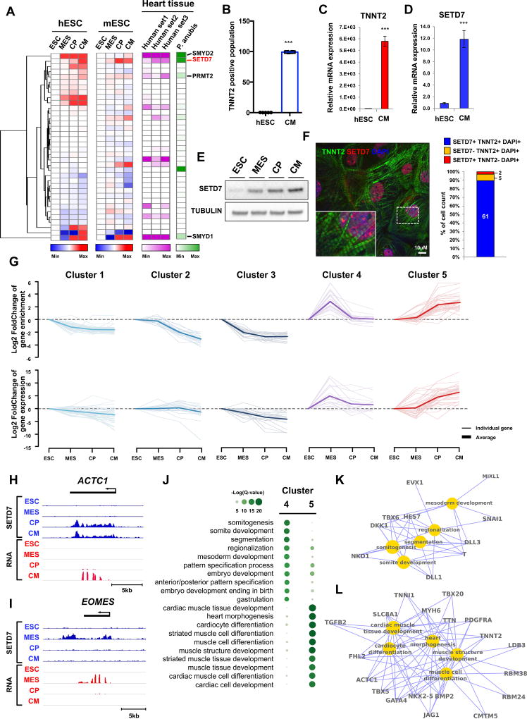
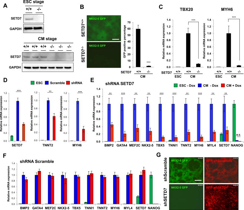
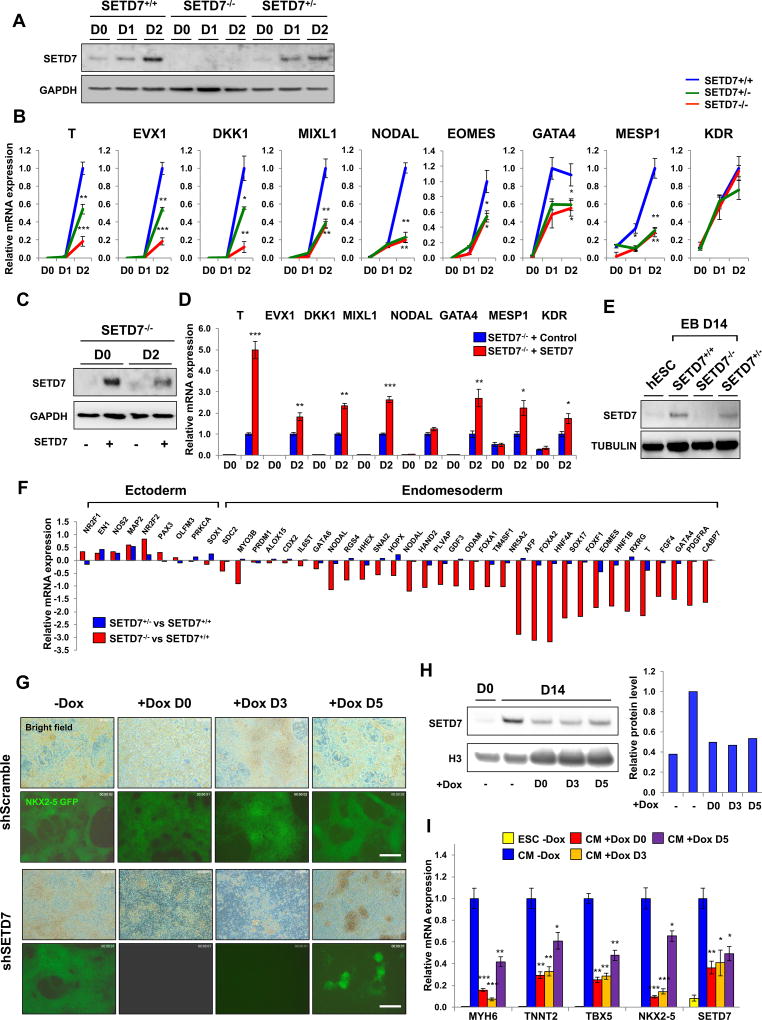
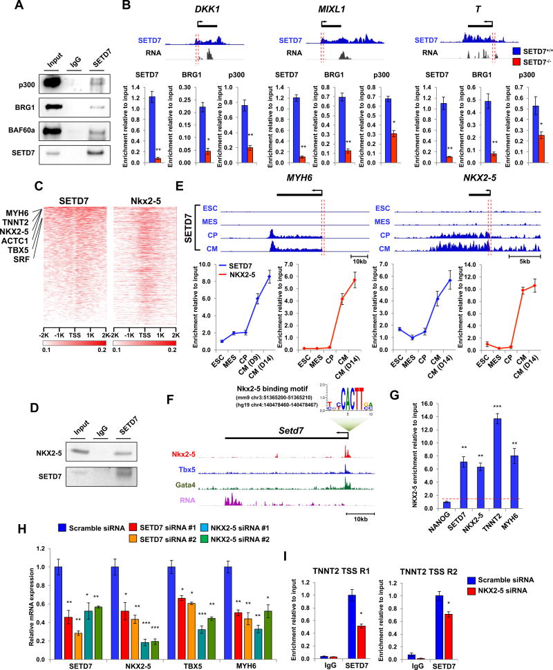
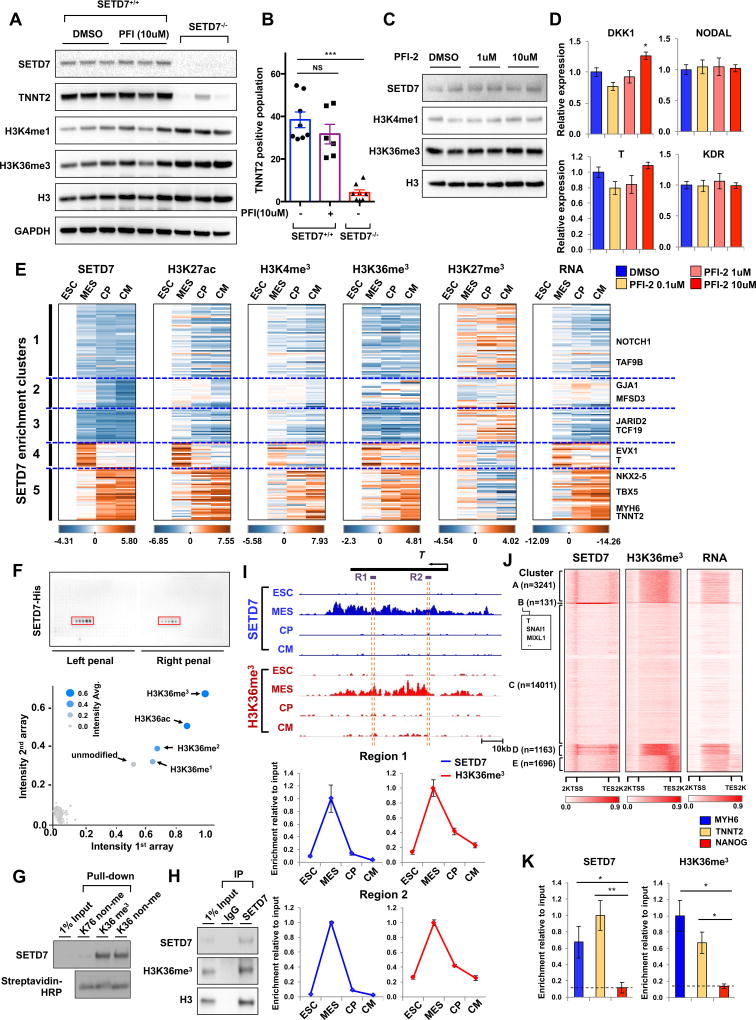
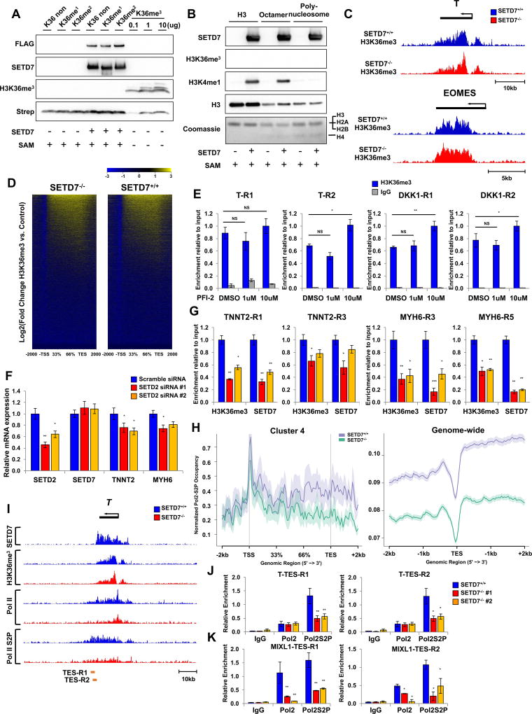
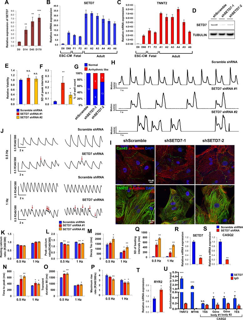
Cardiac development requires coordinated and large-scale rearrangements of the epigenome. The roles and precise mechanisms through which specific epigenetic modifying enzymes control cardiac lineage specification, however, remain unclear. Here we show that the H3K4 methyltransferase SETD7 controls cardiac differentiation by reading H3K36 marks independently of its enzymatic activity. Through chromatin immunoprecipitation sequencing (ChIP-seq), we found that SETD7 targets distinct sets of genes to drive their stage-specific expression during cardiomyocyte differentiation. SETD7 associates with different co-factors at these stages, including SWI/SNF chromatin-remodeling factors during mesodermal formation and the transcription factor NKX2.5 in cardiac progenitors to drive their differentiation. Further analyses revealed that SETD7 binds methylated H3K36 in the bodies of its target genes to facilitate RNA polymerase II (Pol II)-dependent transcription. Moreover, abnormal SETD7 expression impairs functional attributes of terminally differentiated cardiomyocytes. Together, these results reveal how SETD7 acts at sequential steps in cardiac lineage commitment, and they provide insights into crosstalk between dynamic epigenetic marks and chromatin-modifying enzymes.

Keywords: H3K36 methylation; SETD7; cardiomyocyte; epigenetics; histone modification; lineage commitment; stem cell; transcriptional regulation.


Figures

Figure 1
Figure 1. Dynamic target genes of SETD7 during hESC-CM differentiation
(A) RNA-seq expression level of histone methyltransferases at each stage of CM differentiation in hESCs (human), mESCs (mouse), and heart tissues of human and olive baboon (Papio anubis). Heart tissue RNA-seq data were acquired from the Expression Atlas database. (B) Flow cytometry analysis of TNNT2+ cells in H7 hESCs and hESC-CMs (mean ± SEM; n=5). (C–D) RT-qPCR of TNNT2 and SETD7 in hESCs and hESC-CMs. *P < 0.05; **P < 0.01 (one-way ANOVA; mean ± SEM; n=4). (E) Immunoblot analysis of cell lysates at indicated differentiation stages. (F) Immuno-staining of TNNT2, SETD7 in CMs (left). Cell counting of SETD7+ and/or TNNT2+ cells from immune-stained images (right). Scale bar, 10 µm. (G) K-means clustering of SETD7 enrichment during CM differentiation. Y-axis indicates log transformed fold-changes based on day 0 enrichment (Upper) or log transformed fold-changes based on RNA expression at day 0 (Lower). (H–I) Genome browser screenshots of ChIP-seq and RNA-seq for ACTC1 and EOMES. (J) GO analysis of Cluster 4 and Cluster 5. Color code indicates negative log transformed multiple testing adjusted P-value. (K–L) Gene-concept network displaying the gene names associated with the top signaling pathways identified in Cluster 4 and Cluster 5. See also Figure S1 and Table S1.
Figure 2
Figure 2. SETD7 is necessary for CM differentiation
(A) Immunoblot analysis of cell lysates from HES3NKX2-5eGFP/w hESC clone transfected with TALEN targeting start codon of SETD7 gene (upper). Immunoblot analysis of cell lysate from SETD7+/+ and SETD7−/− HES3NKX2-5eGFP/w-derived CMs (lower) (n=3). (B) NKX2-5-GFP expression in SETD7+/+ and SETD7−/− HES3NKX2-5eGFP/w-derived CMs (left). Flow cytometry of GFP+ cells (right). Scale bar, 100 µm. (C) RT-qPCR of TBX20 and MYH6 in SETD7+/+ and SETD7−/− HES3NKX2-5eGFP/w-derived CMs. (D) mRNA levels of SETD7, TNNT2, and MYH6 in control and SETD7 shRNA hESC-CMs. hESCs were infected with lentivirus carrying scramble or SETD7 shRNA. (E–F) Expression level of cardiac-related genes in hESCs and hESC-CMs expressing doxycycline-inducible scramble or SETD7 shRNA. (G) Representative fluorescence microscopy images of hESC HES3NKX2-5eGFP/w-derived CMs. Scale bar, 100 µm. Statistical significance obtained by one-way ANOVA. *P < 0.05; **P < 0.01; ***P < 0.005 (mean ± SEM; n=4). See also Figure S2.
Figure 3
Figure 3. SETD7 is required for transcriptional regulation of lineage specific genes during CM differentiation
(A) Immunoblot analysis of cell lysates from days 0, 1 and 2 of CM differentiation of SETD7+/+, SETD7+/− and SETD7−/− hESC lines. (B) RT-qPCR of mesoderm specific genes in mesoderm lineage cells derived from SETD7+/+, SETD7+/− and SETD7−/− hESC lines. (C) Immunoblot analysis of cell lysates from hESCs and mesoderm lineage cells of SETD7−/− lines carrying control or SETD7 overexpression plasmid. Stable cell lines were generated using lentiviral overexpression system. (D) RT-qPCR of mesoderm specific genes in hESCs and mesoderm lineage cells of SETD7−/− lines carrying control or SETD7 overexpression plasmid. (E) Immunoblot analysis of embryoid body cell lysate from SETD7+/+, SETD7+/−, and SETD7−/− hESC lines. α-tubulin was used as a loading control. (F) RT-qPCR of endomesoderm and ectoderm related genes in embryoid bodies from SETD7+/− and SETD7−/− hESC line compared with SETD7+/+. Total mRNA was extracted from day 14 of embryoid body formation. (G) Representative fluorescence microscopy images of eGFP signal of hESC HES3NKX2-5eGFP/w-derived CMs (day 14) Doxycycline was added to cell media on days 0, 3, and 5 of hESC-CM differentiation. Scale bar, 100 µm. (H) Immunoblot analysis of SETD7 protein levels in hESC HES3NKX2-5eGFP/w-derived CMs (day 14) doxycycline-inducible shRNA for SETD7 or scramble (left). Doxycycline was added to cell media on days 0, 3, and 5 of hESC-CM differentiation. Relative signal intensity of SETD7 (right). (I) RT-qPCR of cardiac specific genes, and SETD7 expression levels in hESC HES3NKX2-5eGFP/w-derived CMs (day 14) doxycycline-inducible shRNA for SETD7 or scramble. Statistical significance obtained by one-way ANOVA. *P < 0.05; **P < 0.01; ***P < 0.005 (mean ± SEM; n=3). See also Figure 2, Figure 3, and Table S2.
Figure 4
Figure 4. SETD7 associates with stage specific co-factors for active gene transcription of its target genes
(A) Immunoblot analysis of SETD7 immunoprecipitates (IP line) and cell lysates (Input line) from mesoderm lineage cells derived from hESCs. (B) Genome browser screenshots of SETD7 ChIP-seq and RNA-seq profiles in DKK1, MIXL1 and T genes at the mesodermal stage of CM differentiation (upper). ChIP-qPCR assay of SETD7, BRG1, and p300 at promoter regions of DKK1, MIXL1, and T genes during CM differentiation (lower). The sequence and genomic location of each primer were described in Table S2. (C) Relative enrichment levels of SETD7 (in human) and Nkx2-5 (in mouse) on TSS of ortholog coding genes in CMs. Heatmaps were ranked by SETD7 enrichment. ChIP-seq data adapted from He et al., 2011. (D) Immunoblot analysis of SETD7 immunoprecipitates (IP line) and cell lysates (Input line) from hESC-CMs. (E) Genome browser screenshots of SETD7 ChIP-seq profiles in MYH6 and NKX2-5 regions in four stages of CM differentiation (upper). ChIP-qPCR of SETD7 and NKX2-5 enrichment at promoter regions of MYH6 and NKX2-5 genes during CM differentiation (lower). Orange bar indicates region targeted by qPCR. (F) Genome browser screenshot of Setd7 gene with a conserved NKX2-5 motif in the promoter region. Enrichment of Nkx2-5, Tbx5, and Gata4 on the promotor region of Setd7 promoter in mouse CMs. ChIP-seq data were adapted from Luna-Zurita et al., 2016. (G) ChIP-qPCR of NKX2-5 enrichment at promoter regions of NANOG, SETD7, NKX2-5, TNNT2, and MYH6 in hESC-CMs. (H) RT-qPCR of mRNA expression of SETD7, NKX2-5, TBX5, and MYH6 genes in CMs transfected with two different sets of siRNA for SETD7 and NKX2-5. (I) ChIP-qPCR of SETD7 and IgG at TSS regions of TNNT2 genes in NKX2-5 knock-down CMs. Statistical significance obtained by one-way ANOVA. *P < 0.05; **P < 0.01; ***P < 0.005 (mean ± SEM; n=3). See also Figure S4.
Figure 5
Figure 5. SETD7 is linked with H3K36 methylation on its target genes
(A) Immunoblot analysis of cell lysates from SETD7+/+ and SETD7−/− lines at day 14 of CM differentiation. PFI-2 and DMSO were treated during entire CM differentiation. (B) Flow cytometry of TNNT2+ cells of SETD7+/+ and SETD7+/- at day 14 of CM differentiation. PFI-2 and DMSO were treated during entire differentiation process (mean ± SEM; n=8 for SETD7+/+ with DMSO and SETD7−/−; n=6 for SETD7+/+ with PFI-2). (C) Immunoblot analysis of mesodermal cells during CM differentiation. DMSO or PFI was treated for 2 days. (D) RT-qPCR of DKK1, NODAL, T, and KDR expression levels in mesodermal cells during CM differentiation. DMSO or PFI was treated for two days. (E) Enrichment levels of histone markers and SETD7 at target genes in each of five clusters and of RNA expression. (F) Spot intensities of two peptide chip arrays. Each peptide chip contained 384 different histone modifications incubated with SETD7 protein. Blue dots represent mean signal intensities from two different peptide arrays. (G) Peptide pull-down assay of histone peptides with SETD7 protein. Biotinylated histone peptides were incubated with GST-SETD7 protein and pulled down with streptavidin coated beads. (H) Immunoblot analysis of SETD7 immunoprecipitates (IP line) and cell lysates (Input line) from hESC-CMs. (I) Genome browser screenshots of H3K36me3 and SETD7 ChIP-seq profiles in T at four stages of CM differentiation (upper). ChIP-qPCR assay of H3K36me3 and SETD7 enrichment at the two gene body regions (dashed orange lines) of the T gene in four stages of CM differentiation (lower). (J) Genome-wide heatmaps of SETD7 and H3K36me3 enrichment in target gene bodies. RNA expression of target genes shown in the right-most heatmap. K-means clustering of SETD7 enrichment used to sort heatmaps. (K) ChIP-qPCR for relative enrichment of SETD7 and H3K36me3 on gene body regions of MYH6, TNNT2 and NANOG genes in left ventricular tissue of healthy individuals (n=5). Statistical significance obtained by one-way ANOVA. *P < 0.05; **P < 0.01; ***P < 0.005 (mean ± SEM; n=3 (panel B); n=5 (panel K)). See also Figure S5 and Figure S6.
Figure 6
Figure 6. SETD7 is not necessary for H3K36me3 but required for Pol II-mediated gene transcription
(A) Immunoblot analysis of SETD7 and H3K36me3. In vitro methylation assay performed with histone peptides and active SETD7-FLAG protein. H3K36me3 peptide used as positive control. (B) Immunoblot analysis of SETD7, H3K4me1, and H3K36me3. In vitro methylation assay was performed with histone H3, histone octamer, polynucleosome and active SETD7-FLAG protein. (C) Genome browser screenshots of H3K36me3 ChIP-seq profiles in T and EOMES genes at mesodermal stages of SETD7+/+ and SETD7−/− lines. (D) Genome-wide heatmaps of H3K36me3 enrichment on genebody region in SETD7+/+ and SETD7−/−(E) ChIP-qPCR of H3K36 enrichment on genebody regions of T and DKK1 genes at mesoderm stage. (F) RT-qPCR of SETD2, SETD7, TNNT2, and MYH6 expression levels in SETD2 knock-down CMs. (G) ChIP-qPCR of SETD7 and H3K36 enrichment on genebody regions of TNNT2 and MYH6 genes in SETD2 knock-down CMs. (H) Average profiles of PolII-S2P enrichment in genebody regions of Cluster 4 genes (left) and genome-wide level (right). (I) Genome browser screenshots of SETD7, H3K36me3, Pol II, and PolII-S2P ChIP-seq profiles on Brachyury (T) gene at mesoderm lineage of SETD7+/+ and SETD−/− lines. (J–K) ChIP-qPCR of PolII and PolII-S2P enrichment at TES region of T and DKK1 genes in SETD7+/+ and SETD7−/− lines at mesoderm stage. Statistical significance obtained by one-way ANOVA. *P < 0.05; **P < 0.01 (mean ± SEM; n=3). See also Figure S7.
Figure 7
Figure 7. SETD7 is critical for calcium handling properties of mature cardiomyocytes
(A) SETD7 expression at various days of hESC-CM differentiation (n=4). (B–C) RT-qPCR of SETD7 and TNNT2 expression levels in hESC, hESC-CMs, fetal heart, and adult heart tissues. (D) Immunoblot analysis of SETD7 in control and SETD7 knock-down lines. (E) Cell viability assay of control and SETD7 knock-down lines. (F) Standard deviation of peak intervals of control and SETD7 knock-down lines. Motion vector from movie images was recorded and analyzed with SONY SI8000 cell motion imaging system (n=18). (G) Total number of wells with arrhythmic-beating hESC-CMs from control and SETD7 knock-down lines. (H) Representative traces of the motion velocity of hESC-CMs infected with scramble or SETD7 shRNA. (I) Immuno-staining of Cxn43, α-Actinin and TNNT2 in control and SETD7 knock-down CMs. (J) Representative ratio-metric calcium imaging traces of control and SETD7 knock-down hESC-CMs. (K–Q) Calcium handling parameters of scramble and SETD7 shRNA lines (n>30). (R–T) Relative RNA expression of SETD7 and calcium handling genes in scramble and shRNA SETD7 groups. (U) ChIP-qPCR of SETD7 and IgG enrichment on TNNT2, MYH6, and CASQ2 genes in hESC-CMs (n=3). Statistical significance obtained by one-way ANOVA. *P < 0.05; **P < 0.01; ***P < 0.005 (mean ± SEM).

Comment in

References

    1. Alexander JM, Hota SK, He D, Thomas S, Ho L, Pennacchio LA, Bruneau BG. Brg1 modulates enhancer activation in mesoderm lineage commitment. Development. 2015;142:1418–1430. - PMC - PubMed
    1. Bannister AJ, Schneider R, Myers FA, Thorne AW, Crane-Robinson C, Kouzarides T. Spatial Distribution of Di- and Tri-methyl Lysine 36 of Histone H3 at Active Genes. J. Biol. Chem. 2005;280:17732–17736. - PubMed
    1. Barsyte-Lovejoy D, Li F, Oudhoff MJ, Tatlock JH, Dong A, Zeng H, Wu H, Freeman SA, Schapira M, Senisterra GA, et al. (R)-PFI-2 is a potent and selective inhibitor of SETD7 methyltransferase activity in cells. Proc. Natl. Acad. Sci. 2014;111:12853–12858. - PMC - PubMed
    1. Brien GL, Gambero G, O’Connell DJ, Jerman E, Turner SA, Egan CM, Dunne EJ, Jurgens MC, Wynne K, Piao L, et al. Polycomb PHF19 binds H3K36me3 and recruits PRC2 and demethylase NO66 to embryonic stem cell genes during differentiation. Nat. Struct. Mol. Biol. 2012;19:1273–1281. - PubMed
    1. Burridge PW, Keller G, Gold JD, Wu JC. Production of De Novo cardiomyocytes: human pluripotent stem cell differentiation and direct reprogramming. Cell Stem Cell. 2012;10:16–28. - PMC - PubMed

Publication types