. 2018 Jun:16:129-138.
doi: 10.1016/j.redox.2018.02.022. Epub 2018 Mar 1.

The immunoproteasome subunit LMP10 mediates angiotensin II-induced retinopathy in mice

Affiliations

The immunoproteasome subunit LMP10 mediates angiotensin II-induced retinopathy in mice

Shuai Wang et al. Redox Biol. 2018 Jun.

Erratum in

Abstract

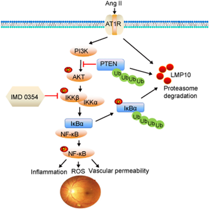
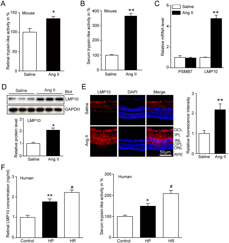
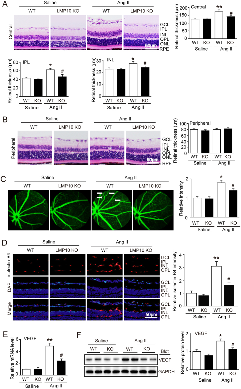
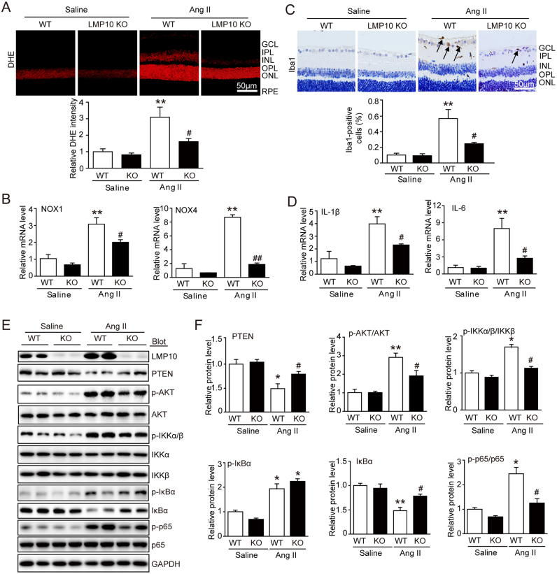
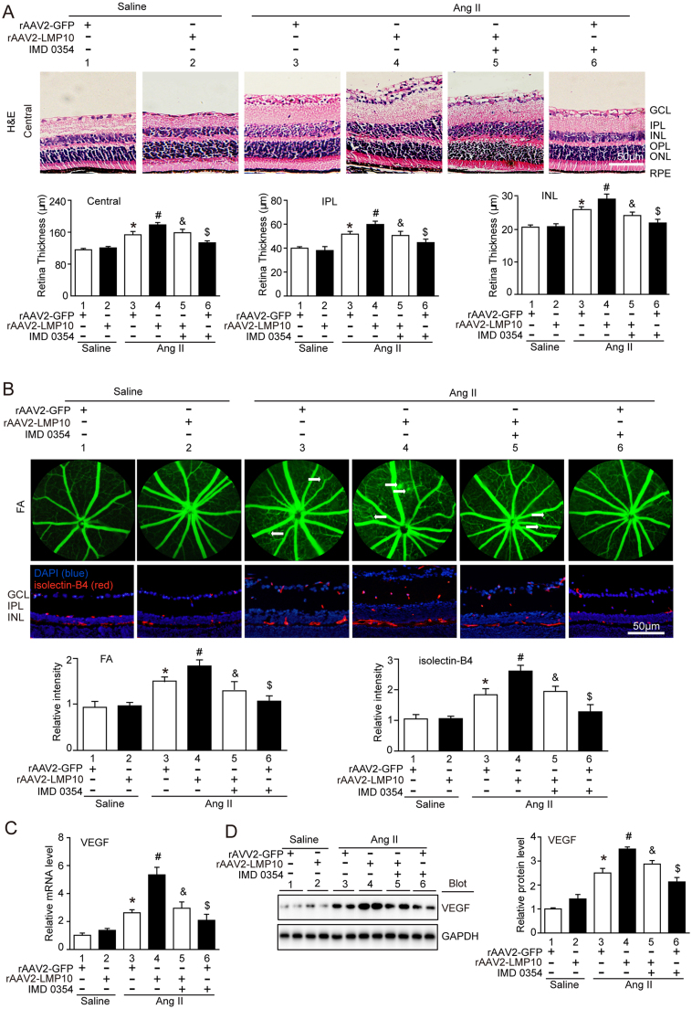
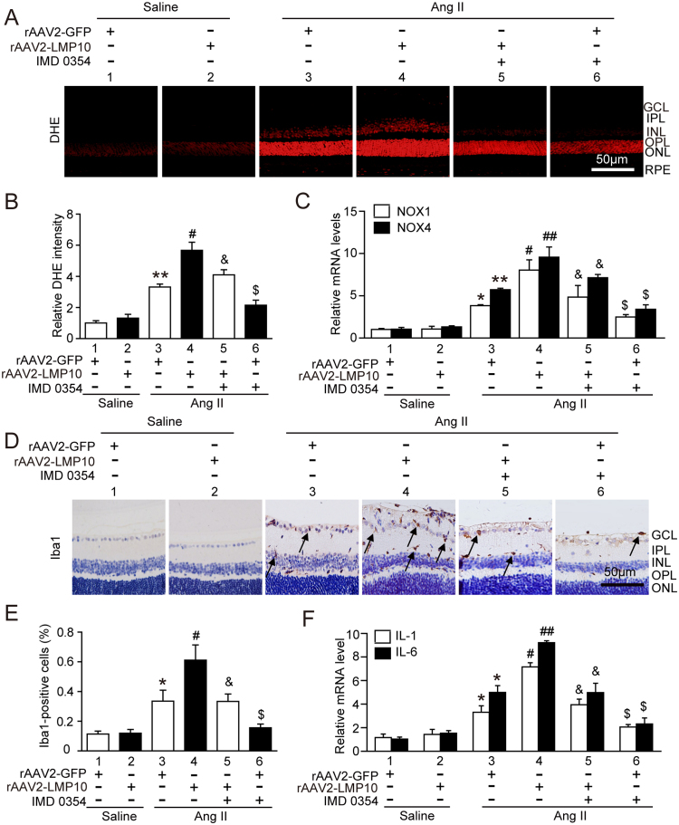
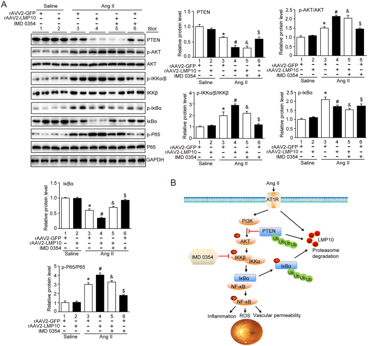
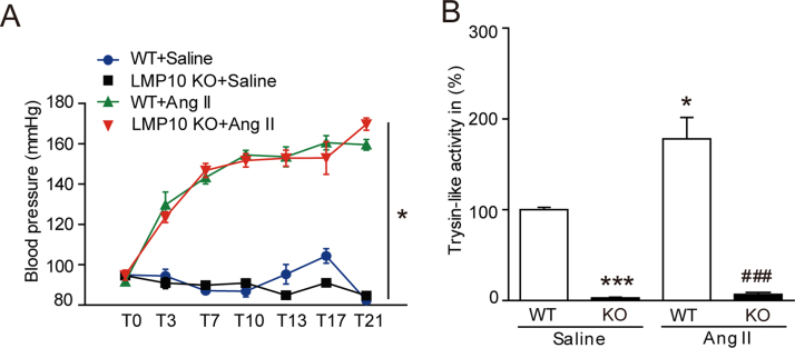
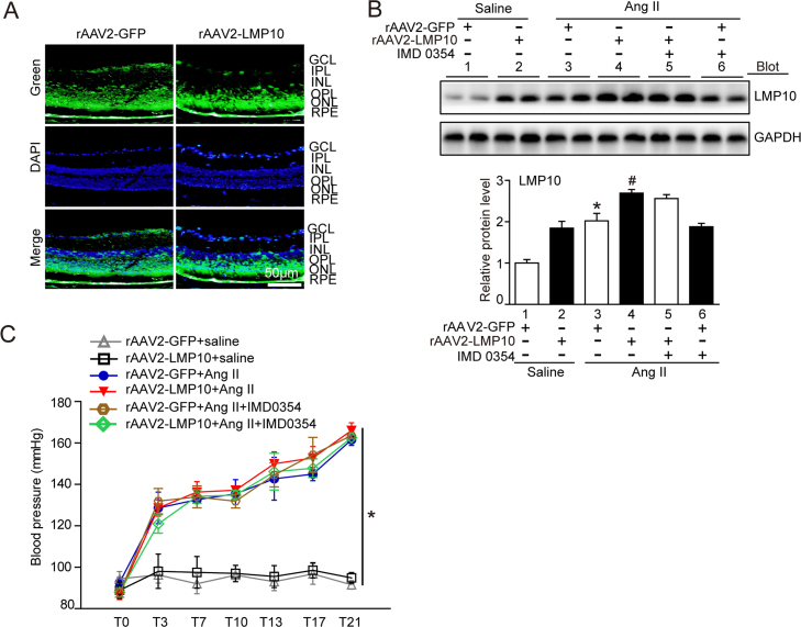
Inflammation has been implicated in a variety of retinal diseases. The immunoproteasome plays a critical role in controlling inflammatory responses, but whether activation of immunoproteasome contributes to angiotensin II (Ang II)-induced retinopathy remains unclear. Hypertensive retinopathy (HR) was induced by infusion of Ang II (3000 ng/kg/min) in wild-type (WT) and immunoproteasome subunit LMP10 knockout (KO) mice for 3 weeks. Changes in retinal morphology, vascular permeability, superoxide production and inflammation were examined by pathological staining. Our results showed that immunoproteasome subunit LMP10 expression and its trypsin-like activity were significantly upregulated in the retinas and serum of Ang II-infused mice and in the serum from patients with hypertensive retinopathy. Moreover, Ang II-infused WT mice showed an increase in the central retinal thickness, vascular permeability, reactive oxygen species (ROS) production and inflammation compared with saline controls, and these effects were significantly attenuated in LMP10 KO mice, but were aggravated in mice intravitreally injected with rAAV2-LMP10. Interestingly, administration of IKKβ specific inhibitor IMD-0354 remarkably blocked an Ang II-induced increase in vascular permeability, oxidative stress and inflammation during retinopathy. Mechanistically, Ang II-induced upregulation of LMP10 promoted PTEN degradation and activation of AKT/IKK signaling, which induced IkBα phosphorylation and subsequent degradation ultimately leading to activation of NF-kB target genes in retinopathy. Therefore, this study provided novel evidence demonstrating that LMP10 is a positive regulator of NF-kB signaling, which contributes to Ang II-induced retinopathy. Strategies for inhibiting LMP10 or IKKβ activity in the eye could serve as a novel therapeutic target for treating hypertensive retinopathy.

Keywords: Angiotensin II; Immunoproteasome LMP10; Inflammation; Oxidative stress; Retinopathy; Vascular permeability.


Figures

fx1
Graphical abstract
Fig. 1
Fig. 1
The trypsin-like activity and LMP10 expression were upregulated in Ang II-infused mice and in hypertensive retinopathy patients. A and B, Measurement of trypsin-like activity in the retinas and serum of wild-type (WT) mice 3 weeks after saline or Ang II infusion (n = 8 mice per group). C, qPCR analysis for the mRNA expression of PSMB7 and LMP10 in the retinas (n = 8 mice per group). D, Immunoblotting analysis of LMP10 (upper) and quantification of the protein bands (lower) (n = 3 mice per group). GAPDH as an internal control. E, Immunostaining of the LMP10 (red) in the retinal sections (left) and quantification of fluorescence intensity (right, n = 4 mice per group). Nuclei were counterstained with DAPI (blue). Scale bar, 50 µm. GCL, ganglion cell layer; IPL, inner plexiform layer; INL, inner nuclear layer; OPL, outer plexiform layer; ONL, outer nuclear layer; RPE, retinal pigment epithelium. F, Measurement of the LMP10 concentration by ELISA assay and the trypsin-like activity in serum of normal controls (n = 31) and hypertension (HP) (n = 31) or hypertensive retinopathy (HR) (n = 30) patients. Data are expressed as the mean ± SEM. *P < 0.05, **P < 0.01 versus saline or normal controls; #P < 0.05 versus hypertension (HP) group.
Fig. 2
Fig. 2
Deficiency of LMP10 attenuates Ang II-induced retinal thickness and vascular permeability. A, Representative H&E staining of central retinal sections and quantification of central retinal thickness of wild-type (WT) and LMP10 knockout (KO) mice 3 weeks after saline or Ang II infusion (n = 10 mice per group). B, Representative H&E staining of peripheral retinal sections (left) and quantification of retinal thickness (n = 10 mice per group). C, Representative angiograms for retinal vessels and quantification of fluorescence intensity. White arrows showed perivascular fluorescein leakage (right, n = 6 mice per group). D, Representative isolectin B4 staining of central retinal sections for endothelial cell proliferation (left) and quantification of fluorescence intensity (right, n = 6 mice per group). Nuclei were counterstained with DAPI (blue). Scale bar, 50 µm. E, qPCR analysis of VEGF mRNA expression in the retinas (n = 6 mice per group). F, Immunoblotting analysis of VEGF expression (left) in the retinas and quantification of protein bands (right) (n = 4 mice per group). GAPDH as an internal control. Data are the mean ± SEM. *P < 0.05, **P < 0.01 versus saline-infused WT mice; #P < 0.05 versus Ang II-infused WT mice. GCL, ganglion cell layer; IPL, inner plexiform layer; INL, inner nuclear layer; OPL, outer plexiform layer; ONL, outer nuclear layer; RPE, retinal pigment epithelium.
Fig. 3
Fig. 3
Knockout of LMP10 reduces Ang II-induced retinal oxidative stress and inflammation. A, Representative dihydroethydium (DHE) staining of central retinal sections (upper) and quantification of DHE intensity (lower) of wild-type (WT) and LMP10 knockout (KO) mice 3 weeks after saline or Ang II infusion (n = 6 mice per group). B, qPCR analysis of the mRNA levels of NOX1 and NOX4 in the retinas (n = 6 mice per group). C, Representative immunohistochemical staining of calcium-binding adapter molecule 1 (Iba1, arrow) in the retinas (upper) and quantification of Iba-positive cells (lower, n = 6 mice per group). Scale bar, 50 µm. D, qPCR analysis of the mRNA levels of IL-1β and IL-6 in the retinas (n = 3 mice per group). E, Immunoblotting analysis of the protein levels of LMP10, PTEN, p-AKT, AKT, p-IKKα/β, IKKα, IKKβ, p-IkBα, IkBα, p-p65 and p65 in the retinas. F, Quantification of the protein bands (n = 4 mice per group). GAPDH as an internal control. Data are the mean ± SEM. *P < 0.05, **P < 0.01 versus saline-infused WT mice; #P < 0.05, ##P < 0.01 versus Ang II-infused WT mice. IL-1β, interleukin-1β; IL-6, interleukin-6; NOX, NADPH oxidase. GCL, ganglion cell layer; IPL, inner plexiform layer; INL, inner nuclear layer; OPL, outer plexiform layer; ONL, outer nuclear layer; RPE, retinal pigment epithelium.
Fig. 4
Fig. 4
Overexpression of LMP10 accelerates, whereas IKKβ inhibitor blunts, Ang II-induced retinal thickness and vascular permeability. A, Representative H&E staining of central retinal sections and quantification of central retinal thickness in WT mice injected with rAAV2-GFP (2.4 × 1012 pfu/ml) or rAAV2-LMP10 (2.4 × 1012 pfu/ml) in the presence or absence of IKKβ inhibitor IMD-0354 (30 mg/kg/d) 3 weeks after saline or Ang II infusion (n = 10 mice per group). B, Representative angiograms for retinal vasculature, isolectin B4 (red) staining of central retinal sections and quantification of fluorescence intensity (right, n = 6 mice per group). White arrows showed perivascular fluorescein leakage. Nuclei were counterstained with DAPI (blue). Scale bar, 50 µm. FA, Fluorescence angiography. C, qPCR analysis of VEGF mRNA levels in the retinas (n = 6 mice per group). D, Immunoblotting analysis of VEGF protein levels in the retinas (left). Quantification of the protein bands (right, n = 4 mice per group). GAPDH as an internal control. Data are the mean ± SEM. *P < 0.05 versus rAAV2-GFP-injected mice infused with saline; #P < 0.05 versus rAAV2-GFP-injected mice infused with Ang II; &P < 0.05 versus rAAV2-LMP10-injected mice infused with Ang II; $P < 0.05 versus rAAV2-GFP-injected mice infused with Ang II.
Fig. 5
Fig. 5
Overexpression of LMP10 aggravates, but IKKβ inhibitor suppresses, Ang II-induced retinal oxidative stress and inflammation. A, Representative dihydroethydium (DHE) staining of central retinal sections in WT mice injected with rAAV2-GFP (2.4 × 1012 pfu/ml) or rAAV2-LMP10 (2.4 × 1012 pfu/ml) in the presence or absence of IKKβ inhibitor IMD-0354 (30 mg/kg/d) 3 weeks after saline or Ang II infusion. Scale bar, 50 µm. B, Quantification of DHE intensity in the retinas (n = 6 mice per group). C, qPCR analysis of the mRNA levels of NOX1 and NOX4 in the retinas (n = 6 mice per group). D, Representative immunohistochemical staining of Iba1 (arrow) in the retinas. Scale bar, 50 µm. E, Quantification of Iba1-positive cells (n = 6 mice per group). F, qPCR analysis of the mRNA levels of IL-1β and IL-6 in the retinas (n = 6 mice per group). Data are the mean ± SEM. *P < 0.05, **P < 0.01 versus rAAV2-GFP-injected mice infused with saline; #P < 0.05, ##P < 0.01 versus rAAV2-GFP-injected mice infused with Ang II; &P < 0.05 versus rAAV2-LMP10-injected mice infused with Ang II; $P < 0.05 versus rAAV2-GFP-injected mice infused with Ang II.
Fig. 6
Fig. 6
Effect of Overexpression of LMP10 or IKK inhibitor on PTEN-AKT and IKK/IkBα/NF-kB signals in the retinas after Ang II infusion. A, Immunoblotting analysis of the protein levels of PTEN, p-AKT, AKT, p-IKKα/β, IKKβ, p-IkBα, IkBα, p-p65 and p65 in the retinas and quantification of the protein bands (n = 4 mice per group). GAPDH as an internal control. Data are the mean ± SEM. *P < 0.05 versus rAAV2-GFP-injected mice infused with saline; #P < 0.05 versus rAAV2-GFP-injected mice infused with Ang II; &P < 0.05 versus rAAV2-LMP10-injected mice infused with Ang II; $P < 0.05 versus rAAV2-GFP-injected mice infused with Ang II. B, A working model for LMP10 to regulate Ang II-induced signaling pathways and retinopathy. In response to Ang II, increased LMP10 promotes PTEN degradation and activation of AKT/IKK signaling, which induces phosphorylation and subsequent degradation of IkBα, ultimately resulting in activation of NF-kB targets and retinopathy.
Fig. S1
Fig. S1
Effect of LMP10 knockout on blood pressure and trypsin-like activity. A, Measurement of systolic blood pressure in wild-type (WT) mice and LMP10 knockout (KO) mice at 3 weeks after Ang II infusion by the tail-cuff method (n = 10 mice per group). B, Proteasome trypsin-like activity in the retinas from WT and LMP10 KO mice after saline or Ang II infusion (n = 10 mice per group). Data are the mean ± SEM. *P < 0.05, * **P < 0.001 versus WT mice infused with saline, ###P < 0.001 versus WT mice infused with Ang II.
Fig. S2
Fig. S2
Infection efficiency of rAAV2 and its effect on blood pressure. A, Infection efficiency of rAAV2-GFP and rAAV2-LMP10 in the retinas indicated by GFP fluorescence (green). Nuclei were counterstained with DAPI (blue). Scale bar, 50 µm. GCL, ganglion cell layer; IPL, inner plexiform layer; INL, inner nuclear layer; OPL, outer plexiform layer; ONL, outer nuclear layer; RPE, retinal pigment epithelium. B, Immunoblotting analysis of LMP10 level in each group (upper). Quantitative analysis of protein intensity (lower, n = 4 mice per group). C, Measurement of systolic blood pressure in rAAV2-GFP- and rAAV2-LMP10-injected mice with or without IMD-0354 injection 3 weeks after Ang II infusion by the tail-cuff method (n = 10 mice per group). Data expressed as the mean ± SEM. *P < 0.05 vs rAAV2-GFP-injected mice infused with saline, #P < 0.05 vs rAAV2-GFP-injected mice infused with Ang II.

References

    1. Katsi V., Marketou M., Vlachopoulos C. Impact of arterial hypertension on the eye. Curr. Hypertens. Rep. 2012;14:581–590. - PubMed
    1. Wong T.Y., McIntosh R. Hypertensive retinopathy signs as risk indicators of cardiovascular morbidity and mortality. Br. Med. Bull. 2005;73–74:57–70. - PubMed
    1. Wilkinson-Berka J.L. Angiotensin and diabetic retinopathy. Int. J. Biochem. Cell Biol. 2006;38:752–765. - PubMed
    1. Verma A., Shan Z., Lei B. ACE2 and Ang-(1-7) confer protection against development of diabetic retinopathy. Mol. Ther. 2012;20:28–36. - PMC - PubMed
    1. Louie J.L., Kapphahn R.J., Ferrington D.A. Proteasome function and protein oxidation in the aged retina. Exp. Eye Res. 2002;75:271–284. - PubMed

Publication types

MeSH terms